Features
A hydatidiform mole is an abnormal pregnancy, in which there is hyperplastic development of the placenta, which often develops into a mass of small vesicles or cysts. The name “hydatidiform” refers to its appearance as a “bunch of grapes”. In a complete hydatidiform mole, there typically is no embryo or fetus by the time it is clinically diagnosed, but in a partial hydatidiform mole there may be an embryo or fetus.
The most common form, complete hydatidiform moles (CHM) are usually sporadic. Interestingly, although these sporadic CHM have a normal number of chromosomes, the DNA that makes up the genetic information in the chromosomes of sporadic CHM is entirely paternally derived, without any maternally inherited DNA. Normally, we have 23 pairs or chromosomes or 46 chromosomes total, with one member of each pair of chromosomes inherited from the mother and one member of each pair inherited from the father.
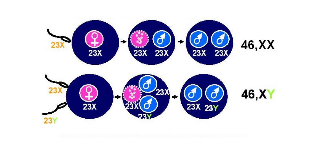
However, in CHM there are also 46 or 23 pairs of chromosomes, but all are paternally inherited, resulting in their designation as “androgenetic” CHM. It is thought that in most CHM this occurs because the nucleus and the 23 chromosomes from the egg become inactive or disappear, while the 23 chromosomes coming from the fertilizing sperm duplicate to make 46 chromosomes.
Another possibility is that such an “empty” egg is fertilized by two sperm, each with 23 chromosomes. (see Figure below). A partial hydatidiform mole (PHM) has 69 chromosomes, with three copies of each chromosome, wherein one copy is maternally inherited and two copies are paternally inherited. In these pregnancies there often is a fetus, but it usually has congenital malformations. This suggests that although identical in sequence, the maternally and paternally inherited member of each pair of chromosomes contribute differently to the developing fetus and placenta. It implies that the abnormal expression of imprinted genes, which are usually only expressed from either the maternally or paternally inherited chromosome is at the origin of the abnormalities seen in androgenetic CHM.

Figure 1: Illustration showing complete hydatidiform moles.

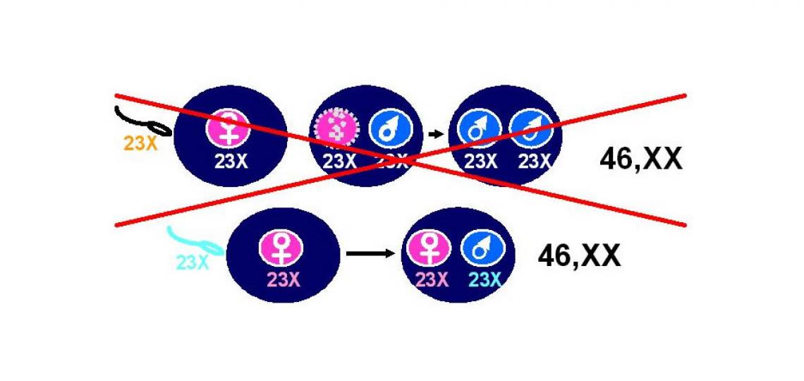
Figure 2: Illustration showing recurrent familial HM are different.
Research Update
Our laboratory is most interested in the study of a rare class of highly recurrent hydatidiform moles. These are usually CHM, although possibly sometimes PHM, that are indistinguishable from androgenetic sporadic CHM pathologically, but are quite different genetically: they have 23 chromosomes inherited from the father and 23 chromosomes inherited from the mother, just like a normally developing pregnancy, leading to the name “biparental” CHM (see Figure 2 above). It was first found that these pregnancies have evidence of abnormal regulation and expression of imprinted genes.
Subsequently it was discovered that women who have these recurrent biparental CHMs have autosomal recessive mutations in a gene named NLRP7, located on chromosome 19 (refs are Murdoch et al, 2006 and Kou et al, 2008). It is believed that a general disruption in the regulation of genetic imprinting, either at the level of establishing imprinting marks in the egg or maintaining imprinting marks in the developing embryo and placenta is at the origin of the defects that lead to the biparental CHM. Our research is currently focused on understanding the mechanisms by which mutations in NLRP7 lead to the abnormalities that result in biparental CHM in women who have these mutations and how this may relate to mechanisms of reproductive loss in general. Another focus of our research on CHM focuses on the discovery of new imprinted genes by studying androgenetic CHM.


