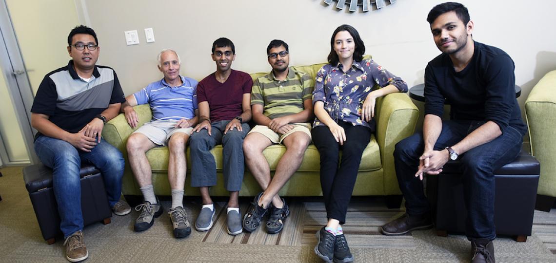
About Our Lab
In our lab, we develop and utilize sophisticated magnetic resonance imaging (MRI) methods to study a variety of problems in neuroscience. A primary focus of the lab is to understand the basic physics and physiology of the functional MRI signal: Blood Oxygen-Level Dependent (BOLD) contrast. We also have particular interests in questions relating to human sensory perception and attention, addressed using high-resolution MRI methods

Superior Colliculus Retinotopy
Images show how visual stimulation and covert visual attention are similarly represented in the superficial layer of human superior colliculus.

Modeling the BOLD Response
New Arterial Impulse Model describes the sequence of events, both physiology and physics, that give rise to the signal used in fMRI.
Theoretical Basis of fMRI Signals
Our main focus concerns the origin of fMRI signals, which are mediated by a combination of neurovascular and neurometabolic coupling. Our laboratory has developed a detailed mathematical model for the fMRI signal based on fluid mechanics and oxygen transport. The results provide excellent descriptions of both oxygen measurements within activated brain tissue, and of the fMRI response evoked by the activation. The models are presently being expanded to provide a detailed picture of oxygen transport throughout the neuropil of cerebral cortex.
We are also interested in the use of fMRI to characterize human spatial vision. To this end, we have developed a powerful new method that uses computed tomography to directly and non-invasively characterize receptive fields in early visual cortex. We aim to utilize these methods to determine how top-down signals such as visual attention affect spatial vision.
Sensation and Attention in the Human Brainstem
Another line of investigation concerns the use of high-resolution structural and functional MRI methods to quantify and visualize functional activity in human brainstem. One set of experiments concern the processing of both bottom-up and top-down signals in the human superior colliculus (SC), a tiny structure that forms millimeter-scale functional maps of visual, auditory, and somatosensory inputs. SC is located near the top of the human brainstem. One goal is to examine how top-down attention affects the nature and spatial distribution of these signals. A second set of experiments have delineated how eye movements are represented in SC. Finally, a new set of experiments examine activity in a different part of brainstem, the vestibular nuclei associated with processing the sense of balance mediated by the semi-circular canals. This work is aimed at understanding the mechanisms of spasticity induced by stroke or other disease. Part of this brainstem research aims to develop new MRI methods that optimize functional data acquisition in this hard-to-image brain region.
Publications
C. A. Greene, S. O. Dumoulin, B. Harvey, D. Ress, Measurement of human visual population receptive fields using back-projection tomography, Journal of Vision 14 , doi: 10.1167/14.1.17 (2014).
S. Katyal, D. Ress, Endogenous attention signals evoked by threshold contrast detection in human superior colliculus, J. Neurosci. 34:3, 892—900 (2014).
D. Ress, B. Chandresekaran, Tonotopic organization in the depth of human inferior colliculus, Front Hum Neurosci 7:586, 1 (2013).
J. H. Kim, R. Khan, J. K. Thompson, D. Ress, Model of the transient neurovascular response based on prompt arterial dilation, J Cereb Blood Flow Metab 33, 1429 (2013).
R. Khan, Q. Zhang, S. Darayan, S. Dhandapani, S. Katyal, C. Greene, C. Bajaj, D. Ress, Surface-based analysis methods for high-resolution functional magnetic resonance imaging, Graphical Models 73, 313 (2011).
S. Katyal, S. Zughni, C.G. Greene, D. Ress, Topography of visual attention and stimulation in human superior colliculus, J Neurophysiol 104:6, 3074 (2010).
D. Ress, J. K. Thompson, B. Rokers, R. K. Khan, A. C. Huk, A model for the transient delivery of oxygen to cerebral cortex, Front Neuroenergetics 1:3, 1—12 (2009).
D. Ress, G. H. Glover, J. Liu, and B. Wandell, Laminar profiles of functional activity in the human brain, Neuroimage 34, 74 (2007).
D. Ress and D. J. Heeger, Neuronal Correlates of Perception in Early Visual Cortex, Nature Neurosci. 6, 414 (2003).
D. J. Heeger and D. Ress, What does fMRI Activity Tell Us about Neuronal Activity? Nature Rev. Neurosci. 3, 142 (2002).
M. L. Harlow, D. Ress, A. Stoschek, R.M. Marshall, and U. J. McMahan, The Architecture of Active Zone Material at the Frog’s Neuromuscular Junction, Nature 409, 479 (2001).
D. Ress, B. T. Backus, D. J. Heeger, Activity in Primary Visual Cortex Predicts Performance in a Visual Detection Task, Nature Neurosci 3, 940 (2000).
G. H. Glover, T. Q. Li, D. Ress, Image-based method for retrospective correction of physiological motion effects in fMRI: RETROICOR. Magn Reson Med 44, 162 (2000).
D. Ress, L. B. DaSilva, et al., Measurement of Laser-Plasma Electron Density with a Soft X-Ray Laser Moiré Deflectometer, Science 265, 514 (1994).


