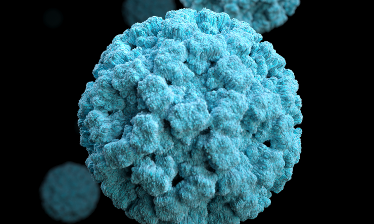
Researchers discovered replication hubs for human norovirus
Human norovirus, a positive-strand RNA virus that is the leading cause of viral gastroenteritis accounting for an estimated 685 million cases and approximately 212,000 deaths globally per year, has no approved vaccines or antivirals. Paving the way for improved drug therapies, researchers at Baylor College of Medicine and the University of Texas, MD Anderson Cancer Center report in Science Advances the discovery of replication hubs for human norovirus, which could lead to designing antiviral drugs to prevent, control or treat these infections.
“When viruses infect cells, they usually create specialized compartments – replication factories – where they form new viruses that infect more cells causing the disease,” said first author Dr. Soni Kaundal, postdoctoral associate in the Verna and Marrs McLean Department of Biochemistry and Molecular Pharmacology at Baylor in the lab of Dr. B.V. Venkataram Prasad, corresponding author of the work. “However, little is known about norovirus’s replication factories.”
Increasing evidence shows that some replication factories typically are not separated from their surroundings by a membrane. Instead, they are biomolecular condensates, structures resembling a bubble formed by liquid-liquid phase separation. These condensates selectively incorporate proteins and other materials needed for viral replication. Liquid-like condensates as replication factories have been extensively studied in other viruses, including the rabies and measles viruses. In this study the researchers investigated whether norovirus forms biomolecular condensates that serve as replication hubs.
“We knew that these condensates are often initiated by a single viral protein capable of binding genetic material, having a flexible region and forming oligomers, molecules made of small numbers of repeating units,” Kaundal said.
The team began their investigation by applying bioinformatic analysis to identify norovirus proteins that would present the characteristics most likely leading to the formation of liquid condensates.
“Working with the human norovirus pandemic strain GII.4, the one responsible for causing most cases of gastroenteritis around the world, we found that the RNA-dependent RNA polymerase has the highest propensity to form biomolecular condensates,” Kaundal said. “This protein has a flexible region, can form oligomers, binds RNA, the norovirus’s genetic material, and plays an essential role during viral replication making copies of the viral RNA. All these characteristics prompted us to experimentally test whether the GII.4 RNA polymerase drives the formation of biomolecular condensates conducive to viral replication.”
“Our experimental studies show that GII.4 RNA polymerase indeed forms highly dynamic liquid-like condensates at physiologically relevant conditions in the lab and that the flexible region of this protein is critical for this process,” said Prasad, professor of molecular virology and microbiology and Alvin Romansky Chair in Biochemistry at Baylor. Prasad also is a member of Baylor’s Dan L Duncan Comprehensive Cancer Center. “Furthermore, the condensates are highly dynamic structures: several can merge forming a larger structure or they can divide into smaller ones; they also move inside the cell, exchanging materials with their surroundings.”
Next, the researchers investigated whether these liquid-like condensates are also formed in human norovirus-infected human intestinal cells. Until recently, studying how norovirus replicates inside cells has been difficult because researchers lacked an effective biological system in which to grow the virus in the lab. But in 2016, the lab of Dr. Mary Estes at Baylor and colleagues succeeded at cultivating human norovirus strains in human intestinal enteroid cultures.
Also known as mini-guts, these cultures are a laboratory model of the human gastrointestinal tract that recapitulates its cellular complexity, diversity and physiology. Human enteroids mimic strain-specific host-virus infection patterns, making them an ideal system to dissect human norovirus infection, as in the current study, to identify strain-specific growth requirements and develop and test treatments and vaccines.
“We showed that liquid-like condensates are formed in human norovirus-infected human intestinal enteroid cultures as well as in the HEK293T human cell line grown in the lab. We propose that these condensates are replication hubs for human norovirus, an elegant solution to the puzzling question of how ribosome-assisted translation of the viral genome is segregated from its replication by the viral polymerase in positive-strand RNA viruses,” Prasad said. “Our bioinformatics analysis also showed that the RNA polymerases of almost all the norovirus strains have a high propensity to form these replication factories, suggesting that this may be a common phenomenon of most noroviruses.”
“This is a remarkable paper, and I was glad we could validate the findings in virus-infected cells using our human intestinal enteroids cultivation system for human norovirus,” said Estes, Distinguished Service Professor and Cullen Foundation Endowed Chair of molecular virology and microbiology at Baylor. Estes also is the co-director of the Gastrointestinal Experimental Model Systems core at the Texas Medical Center Digestive Diseases Center and a member of Baylor’s Dan L Duncan Comprehensive Cancer Center.
The findings not only provide new insight into human norovirus replication but also open new targets for designing antivirals for human norovirus infections, which remain a serious threat in children and immunocompromised patients.
Other contributors to this work include Ramakrishnan Anish, B. Vijayalakshmi Ayyar, Sreejesh Shanker, Gundeep Kaur, Sue E. Crawford, Jeroen Pollet and Fabio Stossi. The authors are affiliated with Baylor College of Medicine and the University of Texas, MD Anderson Cancer Center.
Support for this project was provided by NIH grant P01 AI057788, Robert Welch Foundation grant Q1279, the Center for Advanced Microscopy and Image Informatics (Cancer Prevention and Research Institute of Texas (CPRIT) grant RP170719), the Integrated Microscopy Core at Baylor College of Medicine (NIH grants: DK56338, CA125123, ES030285 and S10OD030414), and CPRIT grant RR160029.


 Credit


