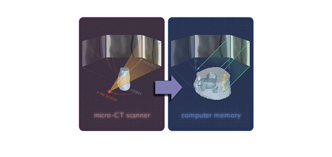
µCT stands for micro-computed tomography – or X-Ray imaging in three dimensions. Similar to hospital CT scanners, these devices are suited for preclinical X-Ray imaging with resolution to the sub-micron scale. Unlike clinical CT scanners where the patient remains still while the scanner images around them, the µCT uses a translatable stage to rotate the sample through hundreds of angular views while the computer computes a stack of virtual cross section slices through the object.

The result is a high-resolution 3d stack with resolution down to 0.5µm for sample sizes up to 75mm. The µCT is a fantastic tool for phenotyping mice, imaging ex-vivo tissue such as lung or even imaging through intact bone.
See our ‘Available Instruments’ list (above right) for details about our μCT scanner.
Optical Imaging & Vital Microscopy Core
Phone: (713) 798-6486
Email: oivm@bcm.edu


