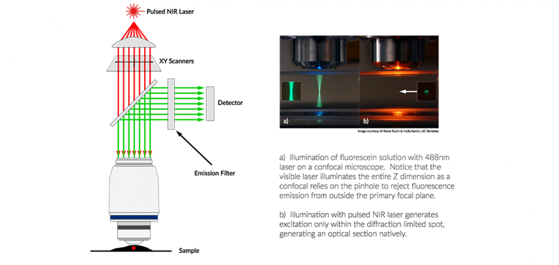
While confocal microscopy uses a pinhole to reject out-of-focus light to generate the optical section, a multi-photon (or 2-photon) microscope uses a pulsed infrared laser to stimulate fluorescence excitation only with the diffraction limited spot of the objective lens, thus creating an inherent optical section.

The advantage of this is two-fold. First, the NIR laser is significantly longer in wavelength than most confocal lasers, so it can penetrate more efficiently through biological tissue. This allows you to use thicker samples than you would with a more traditional confocal microscope. Second, the inherent nature of the optical section generated by photon crowding within the diffraction limited spot allows for direct fluorescence detection without having to travel back through a scanning system. This improves the ability of this system to capture scattered light, which is much more prevalent with thicker samples.
Multi-photon microscopes excel when imaging samples that are thick and opaque. Tissue sections of a few hundred microns or more can be imaged easily with our long working distance objectives. Even more challenging models, such as imaging the intact brain of a live mouse, are made possible with the multi-photon microscope.
See our ‘Available Instruments’ list (above right) for details about our multi-photon microscopes.
Optical Imaging and Vital Microscopy Core
Phone: (713) 798-6486
Email: oivm@bcm.edu


