Organized by Cullen Eye Institute and Department of Ophthalmology, Baylor College of Medicine
The Vision Club Seminar Series provides a platform to connect vision research investigators and clinicians for scientific discussion and research collaboration. The seminar series attracts vision research scientists from the Texas Medical Center as well as in the United States and around the world. The goal of this platform is to develop a rich academic environment, cultivate new scientific ideas and advance the frontier of vision research. Our monthly events are open to all members of vision research community and are generally held at 3 – 4 p.m. (unless otherwise specified) on the second Tuesday of every month.
Seminar Speakers 2022
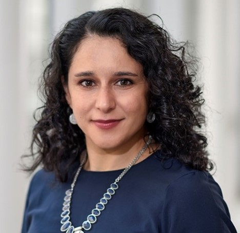
Seminar Title: Elucidating the molecular mechanisms of neural circuit assembly
Time: 3 p.m. via Zoom
Speaker Bio: Dr. Elizabeth Zuniga-Sanchez is an assistant professor in the Department of Ophthalmology at Baylor College of Medicine. She completed her postdoctoral studies in the Howard Hughes Medical Institute lab of Dr. Larry Zipursky at UCLA where she was one of the first to work on mouse development. In a joint collaboration with the lab of Joshua Sanes from Harvard, they combined two transformational technologies, RNA sequencing of different neuronal subclasses and a rapid CRISPR/Cas9-based genetic scheme to test function of specific genes. Using this approach, they uncovered a new signaling pathway responsible for proper synaptic layer formation. As a postdoctoral fellow, Dr. Zuniga-Sanchez was selected as a Helen Hay Whitney fellow and awarded a K99/R00 Pathway to Independence grant. In her independent research, Dr. Zuniga-Sanchez continues to elucidate novel molecular pathways in neural circuit formation using the mouse retina as a model system. Her research is currently funded by a career development award from Research to Prevent Blindness (RPB), an ARVO Genentech Career Development Award for Underrepresented Minority Emerging Vision Scientists, and an R01 from the National Eye Institute.
Seminar Summary: During the development of the nervous system, a myriad of neuronal cell types assemble into a network to form highly stereotypic patterns of connections. How the dendrites and axons from different neuronal subtypes discriminate between one another to form precise and highly reproducible patterns of synaptic connections remains a central issue in neuroscience. Our research focuses on identifying the molecular basis of neural circuit assembly in the developing vertebrate nervous system. We use single cell sequencing, CRISPR technology, live imaging, and single cell labeling to identify the factors that mediate proper connectivity using the mouse retina as a model system. Through these studies, we aim to uncover general principles of neural circuit formation in the developing mammalian nervous system.

Oct. 11, 2022: Stephen Pflugfelder, M.D.
Seminar Title: Retinoid modulation of ocular surface inflammation
Time: 3 pm via Zoom
Speaker Bio: Stephen C. Pflugfelder, M.D., is professor and holder of the James and Margaret Elkins Chair and director of the Ocular Surface Center in the department of ophthalmology at Baylor College of Medicine. He specializes in cornea, ocular surface and tear disorders. Dr. Pflugfelder’s research interests include pathogenesis of keratoconjunctivitis sicca and desiccation-induced inflammation and autoimmunity on the ocular surface. He has published more than 340 peer-reviewed articles and numerous book chapters. He is past president of the International Ocular Surface Society and has served on the editorial boards of Investigative Ophthalmology and Visual Science (associate editor), American Journal of Ophthalmology (associate editor), Scientific Reports, Cornea and The Ocular Surface. He served on the ARVO board as Cornea Trustee and President. He is a member of the Department of Defense Vision Research Panel.
Seminar Summary: Dry eye stress stimulates innate inflammatory pathways in ocular surface epithelial and immune cells. Myeloid cells are recruited to the conjunctiva that produce inflammatory mediators that sensitize nociceptors and stimulate IL-17 production by gamma and delta T cells. IL-17 promotes corneal barrier disruption and conjunctival cornification and goblet cell loss. Retinoid signaling through RXR-alpha suppresses myeloid and gamma delta T cell activation and IL-17 production. Conjunctival and lacrimal gland disease in dry eye can disturb the retinoid axis. The RXR-alpha pathway can be leveraged to treat dry eye inflammation.
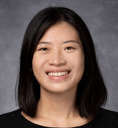
Seminar Title: Light-induced neuronal activity drives the initiation of optic pathway glioma in NF1
Time: 3 p.m. via Zoom
Speaker Bio: Dr. Yuan Pan is an assistant professor in the Department of Symptom Research at the University of Texas MD Anderson Cancer Center. Dr. Pan received her Ph.D. degree from the University of Iowa and her thesis work focused on understanding membrane protein trafficking in photoreceptors. As a postdoctoral fellow, Dr. Pan is co-mentored by Dr. David Gutmann (Washington University) and Michelle Monje (Stanford University) and her research focused on optic pathway gliomas. Leveraging genetically engineered mouse models and neuromodulatory approaches, Dr. Pan’s group is currently working on understanding neuron-glia and neuron-cancer interactions, with a focus on cancer predisposition syndromes such as neurofibromatosis type 1 (NF1).
Seminar Summary: Neurons have recently emerged as key players in driving cancer pathogenesis. While the role of neuronal activity in tumor growth is established, the importance of neuronal activity to tumor initiation was less clear. Individuals with the NF1 cancer predisposition syndrome often develop tumors in the optic pathway (optic glioma) during early childhood, raising the possibility that postnatal light-induced optic nerve neuronal activity drives tumor initiation. In this seminar, Dr. Pan will present how she and her colleagues leveraged a unique mouse model of Nf1 optic glioma to identify how light-induced neuronal activity drives tumorigenesis in the optic nerve.
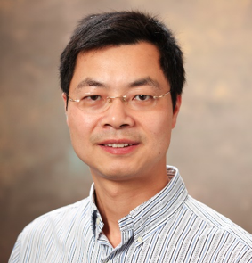
Aug. 9, 2022: Bo Chen, Ph.D.
Seminar Title: Protecting Ganglion Cells and Reprogramming Müller Glia in the Retina
Time: 3 p.m. via Zoom
Speaker Bio: Dr. Bo Chen is professor of ophthalmology and neuroscience at Icahn School of Medicine at Mount Sinai. He received his Ph.D. in pharmacology at the University of Miami School of Medicine, and later pursued postdoctoral training in the Department of Genetics at Harvard University. He was previously an assistant and associate professor in the Departments of Ophthalmology and Neuroscience at Yale University School of Medicine, and is currently a professor in the Departments of Ophthalmology and Neuroscience at the Icahn School of Medicine at Mount Sinai. He received the Karl Kirchgessner Foundation Award for Retinal Research and was a Pew Scholar in the Biomedical Sciences from 2013-2017. He is a reviewer for numerous publications including, but not limited, to Science, Neuron, eLife, PNAS, Cell Reports, Science Advances, and Journal of Neuroscience. Dr. Bo Chen’s research focuses on mechanistic and therapeutic studies of retinal degenerative diseases caused by loss of photoreceptors or retinal ganglion cells, such as age-related macular degeneration, retinitis pigmentosa, and glaucoma. To study these conditions, his laboratory pursues two main strategies: neuroprotective strategy to save existing retinal neurons and neural regenerative strategy to produce new retinal neurons.
Seminar Summary: Retinal ganglion cells (RGCs) and photoreceptors are particularly vulnerable in major retinal degenerative diseases leading to vision impairment and blindness. We will present our recent research progress, as well as future challenges, in protecting RGCs for vision preservation and regenerating photoreceptors by reprogramming Müller glia for vision restoration. Using multiple injury and disease models, we will examine several signaling pathways and transcription factors that may play an important role in the survival and regeneration of RGCs and photoreceptors.
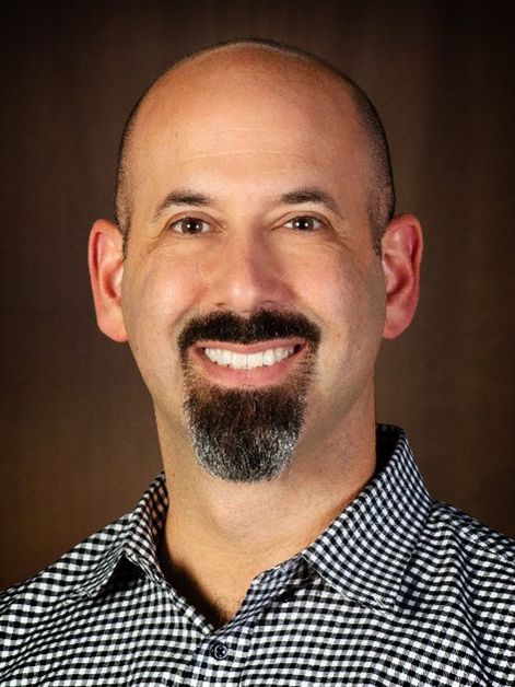
Seminar Title: Glaucoma in mice: new tools, new insights, new questions
Time: 3 p.m. via Zoom
Speaker Bio: Benjamin J. Frankfort, M.D., Ph.D., is an Associate Professor in the Departments of Ophthalmology and Neuroscience at Baylor College of Medicine. He was an undergraduate at Duke University and received his M.D. and Ph.D. degrees from Baylor College of Medicine. He completed a residency in Ophthalmology at the Wilmer Eye Institute of the Johns Hopkins School of Medicine and a fellowship in glaucoma surgery at the Cullen Eye Institute of Baylor College of Medicine. He continued his research training as a K08 recipient under the supervision of Dr. Sam Wu at Baylor College of Medicine. Dr. Frankfort’s research focuses on the impact of eye pressure changes on the visual system. His lab uses a variety of techniques to assess anatomic, physiologic, and molecular changes in injured rodent tissue with resolution ranging from single cells to whole organs. Dr. Frankfort currently holds an R01 and R21 from the National Eye Institute and maintains funding from several foundations. As faculty at Baylor College of Medicine, Dr. Frankfort is highly active in the education of medical students, graduate students, residents, clinical fellows and post-doctoral fellows, and is the Co-Director of the Medical Scientist Training Program (MSTP).
Seminar Summary: Glaucoma is a progressive neurodegenerative disease of retinal ganglion cells (RGCs) and the optic nerve. Despite its wide prevalence and importance as a major public health issue, the earliest changes that occur in glaucoma remain a mystery, hindering early diagnosis and treatment options. In the later stages of glaucoma, RGC regeneration, optic nerve regrowth, and vision restoration techniques are in their infancy.
We will present new techniques developed in the Frankfort lab to study both early glaucoma phenotypes and RGC regeneration. Using the bead injection model of intraocular pressure (IOP) elevation in conjunction with a new vascular assessment workflow, we determined that retinal capillary plexi are differentially susceptible to elevated IOP. Using a series of improvements to RGC immunopanning techniques and optimized FACS, we established a novel system of primary RGC isolation from adult mouse retinas with subsequent culture. With it, we observed robust neurite outgrowth which occurs according to several stereotypical stages.
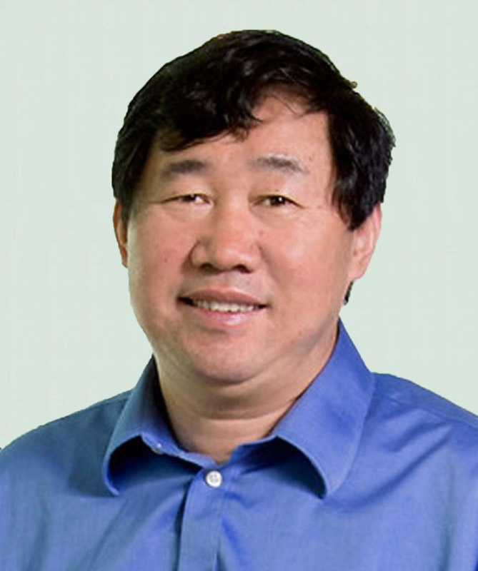
Seminar Title: Deciding a fate: How key transcription factors interact with the epigenetic landscape to promote retinal ganglion cell genesis
Time: 3 p.m. via Zoom
Speaker Bio: Dr. Xiuqian Mu is professor of ophthalmology at the University of Buffalo, Jacobs School of Medicine and Biomedical Sciences. He received his M.D. degree from Qingdao Medical College, China, and his Ph.D. degree in biochemistry and molecular biology from Peking Union Medical College. He received training as a visiting fellow in NIH and postdoctoral associate in MD Anderson Cancer Center, the University of Texas. He was promoted to instructor and assistant professor at MD Anderson Cancer Center and subsequently joined the University of Buffalo as assistant, associate and full professor.
Seminar Summary: My lab is interested in the fundamental mechanisms regulating the shift of cellular states in retinal cell differentiation. One major focus is on how transcription factors influence the epigenetic landscape to promote the genesis of individual retinal cell types such as retinal ganglion cells (RGCs). During development, all retinal cell types emerge from multipotent retinal progenitor cells (RPCs) following distinct trajectories. Recent scRNA-seq studies have further delineated these trajectories, providing a general picture of their relationships and revealing a transitional RPC state shared by all the early lineages. We have now further dissected the changes in the epigenetic landscape along these trajectories by scATAC-seq and identified globally the enhancers, enriched motifs, and potential interacting transcription factors underlying the cell state-specific gene expression in individual lineages. Using CUT&Tag-seq, we identified the enhancers bound directly by four key transcription factors, Otx2, Atoh7, Pou4f2, and Isl1, and uncovered their roles in shaping the epigenetic landscape and controlling the gene expression program in the RGC lineage. Our findings provide experimental support for a general paradigm in which transcription factors collaborate and compete in a sequential and combinatorial fashion to regulate the emergence of distinct retinal cell types such as RGCs from the multipotent RPCs.
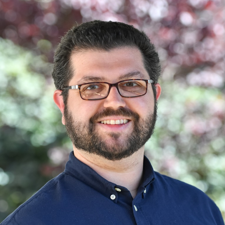
Seminar Title: Selective synaptic disassembly and reassembly of the mature retina in mouse models of glaucoma
Time: 3 p.m. via Zoom
Speaker Bio: Dr. Della Santina is an assistant professor of vision sciences at the University of Houston College of Optometry. He received his Ph.D. in neuroscience from the University of Pisa in Italy studying the functional role of retinal voltage-gated ion channels in processing visual stimuli. He then completed his postdoctoral training at the University of Washington in Seattle under the guidance of Dr. Rachel Wong. His postdoctoral work elucidated the role of neurotransmission in the development of retinal synaptic specificity and identified the relative susceptibility of retinal ganglion cell types to intraocular pressure elevation in mouse models of glaucoma. First at the University of California San Francisco, and now at the University of Houston, his laboratory utilizes electrophysiology, machine learning and large-scale imaging of neural circuits to investigate the effect of retinal neurodegenerative diseases on neural connectivity and functionality.
Seminar Summary: The visual system encodes salient features of the visual scene by employing parallel signal processing pathways. The anatomical basis to such variety starts in the retina with the specific connectivity between types of neurons established through development. The maintenance of this architecture is challenged by neurodegenerative diseases such as glaucoma where post-synaptic neurons, retinal ganglion cells (RGCs) die over time, forcing the existing neural circuits to adapt to a novel environment. We induced RGCs death in mice by experimentally increasing the intraocular pressure and analyzed the intraretinal connectivity of different RGC types with their presynaptic partners, to identify patterns of synaptic rearrangements. We observed that although presynaptic neurons do not die, they lose synaptic ribbons and different RGC types alter their specific connectivity pattern over time and form new connections with former developmental partners. Together, our current data suggests that different neuronal types engage diverse strategies in response to degeneration; leveraging these differences could be the key to develop early functional tests for neurodegenerative diseases.

Seminar Title: Ultrastructural analysis of Cep290 mutant cilia in mouse photoreceptors
Time: 3 p.m. via Zoom
Speaker Bio: Dr. Abigail Moye is a postdoctoral fellow in Dr. Theodore Wensel’s Laboratory, Department of Biochemistry and Molecular Biology, Baylor College of Medicine. Her research is funded by NIH F32 Postdoctoral Fellowship.
Seminar Summary: CEP290 (centrosomal protein 290), a large multi-domain containing protein, is a key component of the transition zone in many primary and motile cilia. Mutations in Cep290 cause several ciliopathies, including Meckel Syndrome (MKS) and retinal specific disorders such as Leber Congenital Amaurosis (LCA). The CEP290 protein has been proposed as a component of the “Y-links” structures, which span from the axoneme microtubule doublets to the ciliary membrane in the connecting cilium (CC) of photoreceptor cells. In this presentation, I will discuss the retinal phenotypes in two Cep290 mutant mouse models, as well as our use of multiple advanced microscopies to determine the location of CEP290 protein and the structural effects resulting from mutations in CEP290 at the nanometer scale.
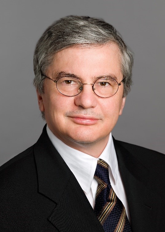
Seminar Title: Identification of Novel Mechanisms of Angiogenesis
Time: 3 p.m. via Zoom
Speaker Bio: Dr. Napoleone Ferrara is distinguished professor of pathology and ophthalmology and Hildyard Endowed chair at UC San Diego, Calif. Before joining UCSD in 2013, he was senior scientist, staff scientist and Genentech fellow at Genentech. At Genentech, he discovered vascular endothelial growth factor (VEGF) and its receptors and developed the first anti-VEGF antibody drug bevacizumab (Avastin) for cancer therapy. His work also led to the development of ranibizumab (Lucentis) for the therapy of ocular vascular diseases. He has received numerous awards, including Lasker-DeBakey Clinical Medical Research Award in 2010 and Breakthrough Prize in Life Sciences in 2013. At UCSD, his research focuses on investigating angiogenic mechanisms in tumor and ocular disease.
Seminar Summary: Dr. Ferrara’s research focuses on investigating mechanisms of angiogenesis in disease conditions alternative to VEGF. He will present his recent findings in therapeutic angiogenesis.

Seminar Title: Top-down feedback controls neural responses in primate visual cortex
Seminar Time: 3 p.m.
Speaker Bio: Dr. Lauri Nurminen is assistant professor of vision science at the University of Houston College of Optometry. He received his Ph.D. degree in neuroscience from the University of Helsinki, Finland. He completed postdoctoral training at the Moran Eye Center of the University of Utah under the guidance of Dr. Alessandra Angelucci. His postdoctoral work elucidated the neural circuit basis of receptive fields of primary visual cortex neurons in non-human primates. Part of his NIH K99 training was completed under the guidance of Dr. John Reynolds at the Salk Institute for Biological Studies where he worked with awake, behaving marmoset monkeys. His laboratory utilizes electrophysiology, optogenetics, and behavioral techniques in awake and behaving marmosets to elucidate the roles of recurrent connections between cortical areas in visual computation and perception.
Seminar Summary: Sensory information travels along feedforward connections through a hierarchy of cortical areas, which, in turn, send feedback connections to lower-order areas. Feedback has been implicated in attention, expectation, and sensory context, but how feedback could implement such diverse functions remains unknown. In my presentation, I will show how we have used optogenetic inactivation of the axon terminals of feedback connections from the second visual area (V2) to the primary visual cortex (V1) in non-human primates (Common Marmoset) to determine how feedback affects neural responses in V1. Our results show that reducing feedback activity increases V1 cells’ receptive field size, decreases their responses to stimuli confined to the RF, and increases their responses to stimuli extending into the proximal surround, therefore reducing surround suppression. Moreover, a stronger reduction of V2 feedback activity leads to a progressive increase in RF size and a decrease in response amplitude. Our results indicate that feedback modulates RF size, surround suppression, and response amplitude, similar to the modulatory effects of visual-spatial attention.
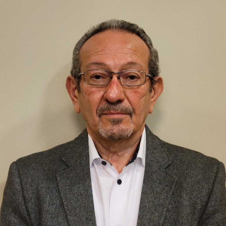
Seminar Title: Role of riboflavin in vision
Seminar Time: 3 p.m. via Zoom
Speaker Bio: Dr. Muayyad Al-Ubaidi is Professor of Biomedical Engineering at Cullen College of Engineering, University of Houston. He is a leader in mouse transgenesis and has been very active in the production of transgenic models of inherited retinal disease.
Seminar Summary: Ariboflavinosis is a pathological condition occurring because of riboflavin (vitamin B2) deficiency. Riboflavin is intracellularly converted to flavin mononucleotide (FMN) and flavin adenine dinucleotide (FAD), both are cofactors for many enzymatic reaction. Ariboflavinosis can be the result of mutation is riboflavin transporters or improper dietary intake of the vitamin. Clinical manifestations of ariboflavinosis include neurological and visual abnormalities. However, the mechanism of the visual defects is not understood. In this presentation, we will address how riboflavin is delivered to the retina, maintained at high level, the mechanism that leads to visual defects resulting from ariboflavinosis and the metabolic changes associated with manipulations in retinal flavin levels.

Seminar Title: Investigating IL-2 Corneal Epithelial Signaling
Seminar Time: 3 p.m. via Zoom
Speaker Bio: Dr. Cintia De Paiva is Associate Professor of Ophthalmology at Baylor College of Medicine. Her research focuses on pathogenesis of dry eye-related diseases with the ultimate goal to improve the diagnosis, prognosis and therapy of dry eye.
Seminar Summary: The cornea is richly innervated and responds to various chemical, mechanical, and thermal stimuli to initiate reflex blinking and tearing. Neuropeptides produced by corneal nerves maintain epithelial integrity and stimulate wound healing. In turn, corneal epithelial cells support the nerves functioning as surrogate glial cells. However, the superficial location of the corneal nerves renders them susceptible to desiccating stress, mechanical trauma, and inflammation that can sensitize them, causing pain and excessive blinking. Diseases that affect corneal nerves such as dry eye and neurotrophic keratitis can lead to debilitating ocular pain, ulceration, and opacification and may require aggressive therapies, including corneal transplantation, to restore vision.
IL-2 is a cytokine with a central role in the immune system, promoting activation, clonal expansion, and deletion of T lymphocytes. IL-2 is detectable in the tears of healthy subjects. IL-2 signals through its heterotrimeric receptor (IL-2R) consisting of the alpha (CD25), beta, and gamma chains. IL-2 binds directly to CD25, making this receptor subunit key to IL-2 signaling. Non-immune CD25 and IL-2Rbeta expression have been reported in several different types of epithelial cells. We and others have reported that the corneal epithelium exhibits strong immunoreactivity to CD25. We have provocative and exciting data suggesting that the corneal CD25 is a functional receptor and that an IL-2 corneal epithelial signaling exists.


