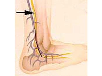
A nerve biopsy involves the process of removing a small length of a peripheral nerve for histologic examination under the microscope. Several nerves can be biopsied but the major requirement for selection of a nerve for biopsy is that it should be a purely sensory nerve. Biopsy of a nerve with any motor function carries the risk of inducing motor deficits that far outweigh the possible diagnostic benefit of the biopsy. The most accessible sensory nerve for biopsy is the sural nerve located behind the ankle. The procedure involves injection of a local anesthetic in the area to be biopsied. An incision about two inches long is then made behind the ankle. The skin is retracted and blunt instruments are used to gently separate the nerve from the adjacent structures, including blood vessels and subcutaneous fatty and fibrous tissues. Once a length of nerve equaling 1½ - 2 inches is freed, the nerve is cut and immediately sent to the laboratory for processing. The superficial peroneal nerve, located at the front of the shin, may also be biopsied.
The discomfort associated with the biopsy procedure related to the injection of local anesthetic is felt as a needle stick and a slight stinging and burning sensation. Other discomfort is related to the cutting of the nerve which usually results in a very transient and sudden sharp pain.
Potential Complications

The major potential complications of a nerve biopsy include bleeding, infection, scarring, permanent numbness on the outer side of the foot, and the possibility of developing chronic pain at the site of the biopsy. Bleeding and infection are the usual potential complications of any surgical procedure. Bleeding is minimal once care is taken to ensure avoidance of "blood thinning" agents (anticoagulants, aspirin, etc.) prior to the procedure. The risk of infection is slightly increased in sural nerve biopsy due to the location of the incision site which is close to the foot. Incision site infections are usually easily treated with oral antibiotics. The sural nerve provides sensation for the outer side of the foot and once it is removed, sensation will most likely be permanently lost, or possibly just altered, on the outer side of the foot. Chronic pain at the site of the nerve biopsy occurs in 3 to 5 percent of patients undergoing a nerve biopsy.
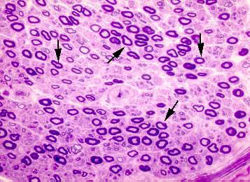
In addition to routine histology, a properly processed nerve biopsy must include semi-thin sections and teased nerve fiber preparations. These techniques are necessary to arrive at a proper diagnostic interpretation of the nerve biopsy.

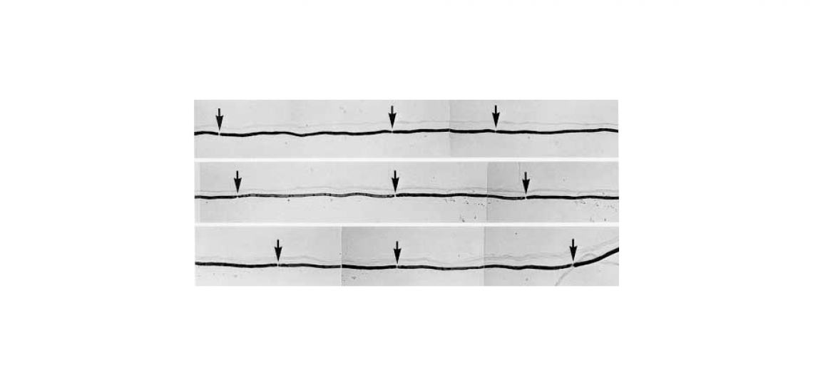
Teased nerve fiber preparation demonstrating the length of nerve consisting of nine consecutive internodes (arrows point to the nodes of Ranvier). There is variation in the length of internodes suggesting a demyelinating-remyelinating process.


