About the MicroCT Lab
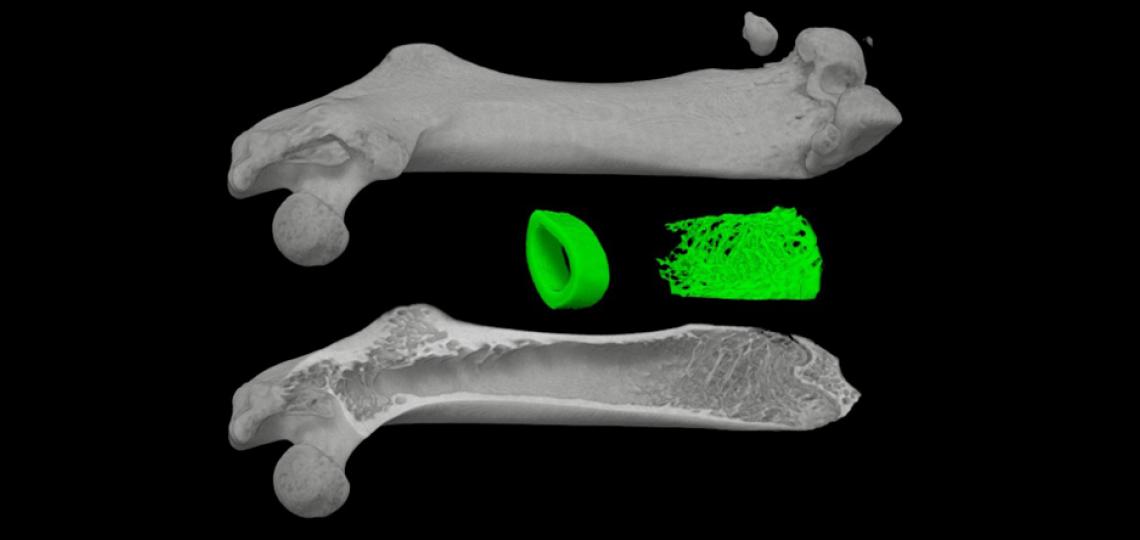
The μCT 40 MicroCT scanner located at Baylor College of Medicine allows for visualization, measurement, and quantification of the structure of bone. This system creates a three-dimensional model from a large number of two-dimensional X-ray images taken around a single axis of rotation.
The high-resolution scanner can capture detail at 6 microns, although a typical scanning resolution of 12 microns is sufficient for most applications. Scans taken of long bones have specific trabecular or cortical analysis performed on them. Spines, skulls, and whole mice skeletons can be scanned and used for phenotypic analysis.
The high resolution, multiple scan nature of the MicroCT scanner requires long scan times, often requiring several hours for the scan and several hours to reconstruct the image from the scan data. A typical scan will be run overnight with analysis to take place the following day. Upon completion of a scan and reconstruction, the samples can be analyzed or simply viewed to examine morphological features.
The standard types of analysis available are cortical and trabecular analysis, yielding the total volume, bone volume, difference, trabecular thickness, number of trabeculae, trabecular separation, connectivity, and other measurements. This scanner functions as part of a bone analysis core.
Sample Preparation
The following is the recommended sample preparation procedure for samples to be scanned by MicroCT.
Collect bone of interest using the standard protocol for bone collection (fixation in formalin for 48 hours, followed by 70 percent ethanol, then store at 4 degrees until you scan). Any other solution that is appropriate for your specimens (e.g. PBS) is good for the scanner as long as it is immersed in a liquid. Plastic embedded specimens can also be scanned but with some limitations. Samples are typically scanned in 70 percent alcohol.
Ideally, the whole femur or tibia is to be collected to study cortical and trabecular bone. Cortical bone analysis is performed on the mid diaphyseal shaft or a region 1-2 mm from the distal or proximal growth plate. So for example, if you have a sample consisting of only a midshaft cut femur with the knee joint, the distal femur can be analyzed. Trabecular bone is also analyzed in similar manner in the distal or proximal end of the bone starting from the end of the growth plate and taking a certain number of cuts (typically 50-150) as a region of analysis. Standard trabecular bone analysis on spine samples can be performed on the L3-L6 regions. Other regions of interest are up to the investigator and the project, but there are no standardized ways to measure other regions for the time being (e.g. calvaria, rib cage, humerus).
Main Bone Parameters
The main bone parameters returned from the analysis are:
- TV: total volume [mm^3]
- BV: bone volume [mm^3]
- BV/TV: the relative volume of calcified tissue in the selected volume of interest (VOI)
- Tb.Th: it measures the thickness of the trabecular structure
- Tb.N: indicates the number of trabeculae
- Tb.Sp: trabecular separation; it is a measurement of the thickness of the spaces between the trabeculae. It is inversely proportional to the trabecular thickness
- Conn.D: connectivity density, 3-D connectivity index; it is a measure of the degree to which a structure is multiply connected
- SMI: structure model index; this measurement is related to the architecture of the structure and ranges between 0 and 3. An SMI of 0 signifies that the structure is only plates, whereas a value of 3 means only cylindrical rods.
Service Requests
Special rates are available for bone investigators and institutional users. Please contact us for pricing.
MicroCT Request Form
Complete form to request services from the MicroCT Lab.


