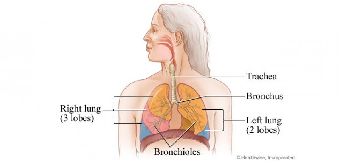
Plication of the diaphragm is performed for paralysis or eventration (abnormal elevation/shape) of the diaphragm which can result in breathing difficulties. Diaphragm paralysis is typically due to damage to the phrenic nerve; eventration is most commonly congenital.
Surgical plication to stabilize the diaphragm is needed to prevent the lungs from ballooning outward during expiration (breathing out), the opposite of normal function.
Evaluation of a patient with diaphragm paralysis includes careful examination, imaging, and respiratory workup. A careful, detailed history of the onset, timing of paralysis, and severity will allow the surgeon to determine the likelihood of spontaneous recovery and the possible cause of the paralysis.
Associated congenital defects
Many candidates for the procedure due to eventration have other congenital disorder such as:
- Undescended testicle
- Abdominal visceral transposition
- Cleft lip and palate
- Hypoplastic arch disorder
- Patent ductus arteriosus
- Ventricular septal defect (VSD)
- Coarctation of the aorta
- Gastric volvulus
- Horseshoe kidney
Surgical repair
There are several plication techniques available. A number of variations exist and are performed by thoracoscopic, laparoscopic, open or hybrid techniques. With improved robotic and videoscopic equipment, the trend towards less invasive procedures is on the rise.
The goal of plication is to tighten the loose diaphragm, making it firm and flat so other breathing muscles can work more effectively.


 Credit
