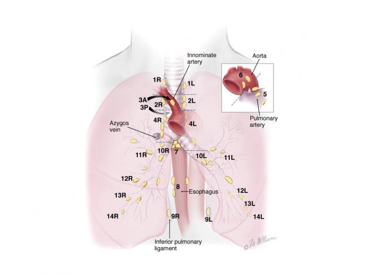
Mediastinal lymph node dissection is performed to assist in the staging and treatment of non-small cell lung cancer.
Anywhere from a dozen to over 40 lymph nodes may be removed and sent to pathology for sampling.
The surgeon will perform either a right- or left-sided lymphadenectomy, sometimes with the use of video-assisted thoracic surgery (VATS).
They first make an incision between the fifth and sixth ribs.
The patient’s lung is retracted and a vein on the back wall of the ribcage by their esophagus (azygous) is exposed. The lymph node packet is removed from the surrounding tissues in this area.
The dissection is continued near the aortic arch, where more lymph nodes are found and removed. Lymph nodes near the trachea and left and right main bronchi, as well as the esophagus, are removed and sampled.
References
Sugarbaker DJ, Bueno R, Krasna MJ, Mentzer SJ, Zellos L (eds). Adult Chest Surgery. New York: The McGraw-Hill Company, 2009.
Mountain CF. A new international staging system for lung cancer. Chest. 89:225–33S, 1986.
Mountain CF, Dresler CM. Regional lymph node classification for lung cancer staging. Chest. 111:1718–23, 1997.
Kaseda S, Hangai N, Yamamoto S, Kitano M. Lobectomy with extended lymph node dissection by video-assisted thoracic surgery for lung cancer. Surg Endosc. 11:703–6, 1997.


 Credit
