


Organized by Cullen Eye Institute and Department of Ophthalmology, Baylor College of Medicine
Sponsored by Retinal Research Foundation
The Vision Club Seminar Series provides a platform to connect vision research investigators and clinicians for scientific discussion and research collaboration. The seminar series attracts vision research scientists from the Texas Medical Center as well as in the United States and around the world. The goal of this platform is to develop a rich academic environment, cultivate new scientific ideas and advance the frontier of vision research. Our monthly events are open to all members of vision research community and are generally held at 3 – 4 p.m. (unless otherwise specified) on the second Tuesday of every month.
Seminar Online Link
Or in-person at Cullen Eye Institute Auditorium, NC202, 6565 Fannin St. Houston
Contact Jessica for any questions.
Seminar Speakers 2025

Dec. 11, 2025: Shiva Swamynathan, Ph.D.
Seminar Title: Corneal Functions of Apicobasal Polarity-Determinant PARD3
Time: 3 p.m. via Zoom and in-person
Speaker Bio: Shiva Swamynathan graduated with a Ph.D. in Life Sciences from the Center for Cellular and Molecular Biology in Hyderabad, India, in 1996. Shiva trained as a post-doctoral fellow with Dr. Joram Piatigorsky at the National Eye Institute, NIH, and worked as an Assistant Professor of Ophthalmology (2007-2015), and Associate Professor of Ophthalmology with Tenure (2015-2023) at the University of Pittsburgh School of Medicine. Shiva joined the Department of Ophthalmology, Morsani College of Medicine, University of South Florida, Tampa in Jan 2023 as a Tenured Professor, where he directs the Ocular Surface Biology Laboratory. Shiva is an Editorial Board Member for 'The Ocular Surface' (2023-Present), a member of the ARVO Professional Development Committee (2024-2027) and a standing member of the Biology and Development of the Eye study section (2024-2028). The research in the Swamynathan laboratory, supported by NIH grants RO1 EY026533 and RO1 EY031684, is focused on elucidating the ocular surface functions of: (i) Krüppel-like transcription factors KLF4 and KLF5; (ii) immunomodulatory molecule SLURP1; and (iii) apicobasal polarity-determinant PARD3.
Seminar Summary: Shiva Swamynathan will discuss the corneal epithelial functions of PARD3, a key determinant of epithelial apicobasal polarity. The Swamynathan lab used a mouse model with cornea-specific ablation of Pard3 and a complementary human corneal epithelial cell culture model with CRISPR-mediated mutation in PARD3 to discover that PARD3 ablation disrupts corneal epithelial apicobasal polarity, barrier function, and actin cytoskeleton, while elevating cell proliferation and expression of EMT transcription factors. These findings will be presented along with more recent results that suggest involvement of PARD3 in regulating corneal sensory innervation and genome stability.

Nov. 13, 2025: Justin Kumar, Ph.D.
Seminar Title: Interconversion of compound eyes and ocelli through the dialing of transcription factor levels
Time: 3 p.m. via Zoom and in-person
Speaker Bio: Dr. Justin P. Kumar is a Professor in the Department of Biology at Indiana University. His laboratory employs the fruit fly Drosophila melanogaster to investigate how transcription factors, particularly the Pax6 proteins Eyeless and Twin of Eyeless, govern the formation, identity, and interconversion of visual organs such as compound eyes and ocelli. His research has revealed how fine-tuned modulation of gene expression within the retinal determination network can reprogram tissue fates, uncovering fundamental principles of developmental biology and the evolutionary diversification of visual systems. Dr. Kumar has been recognized with numerous honors, including election as a Fellow of the American Association for the Advancement of Science (AAAS) and the Indiana University Trustees Teaching Award. He has published more than 70 scientific papers and serves on the editorial boards of Developmental Biology and Developmental Dynamics.
Seminar Summary: The adult visual system of Drosophila melanogaster is composed of two compound eyes, a pair of extra retinal eyelets, and a trio of simple eyes called ocelli. These distinct eye types have evolved independently from each for over 400-500 million years and are anatomically distinct, connect to different regions of the brain, and control unique arrays of visual and circadian behaviors. The fate of compound eyes and ocelli is controlled by the activity of the Paired Box-6 (Pax6) transcription factors Eyeless (Ey) and Twin of Eyeless (Toy). Pax6 plays prominent roles in the developing eye of all seeing animals with strong loss-of-function mutants being associated with microphthalmia or anophthalmia-like phenotypes in humans, mice, zebrafish, and the fruit fly. Pax6 is also characterized by its ability to reprogram the fate of entire cell populations – the forced expression of Ey and Toy can induce the formation of ectopic eyes within non-ocular tissues. Ey and Toy differentially control the fate of the compound eyes and ocelli with the former specifying the identity of the compound eye and the latter directing the fate of the ocelli. These proteins appear to regulate a common set of downstream targets but activate them at vastly different levels within the two visual organs. This is due to Ey having a much stronger transcriptional activation domain than Toy. Quite surprisingly, simply modulating the levels or activity of Ey/Toy downstream targets induces a dramatic reprogramming of the ocelli into compound eyes. One target gene, sine oculis (so), encodes a DNA binding protein that can function as a transcriptional activator when bound to Eyes Absent (Eya) or a repressor when bound to Groucho (Gro). Our findings suggests that the stochiometric balance between So-Eya and So-Gro complexes is critical for selecting between the two different types of visual organ fates. Finally, ratcheting down levels of Ey and Toy (as well as their downstream targets) within the developing compound eye results in the reverse fate change and induces its conversion into ocelli, antennae, maxillary palps, and head epidermis. Our results suggest that adjusting the expression levels of field-level selector genes across a wide threshold range is a viable mechanism for specifying a diversity of cell and organ fates.
Oct. 9, 2025: Felice Dunn, Ph.D.
Seminar Title: Detection of retinal degeneration by measuring reflexive eye movements
Time: 3 p.m. via Zoom and in-person
Speaker Bio: Felice Dunn began her studies in neuroscience as an undergraduate at Brown University where she worked with David Berson in the discovery of intrinsically photosensitive ganglion cells. She then continued her studies in vision at the University of Washington with Fred Rieke as a graduate student and with Rachel Wong as a postdoctoral fellow. With Fred Rieke, she examined retinal gain controls that enable vision across a broad range of mean light levels. With Rachel Wong, she uncovered developmental patterns of synapse formation. In between, she studied the fidelity of olfactory processing at the NIH with Mark Stopfer. Currently at UCSF, her lab examines how plasticity can occur in the adult retina. Her lab has developed tools for structural and functional assessment and a novel method of studying loss of primary sensory receptors in the adult retina. Her lab has pioneered unique tools for photoreceptor perturbation to uncover the possibilities for synaptogenesis and functional compensation that could occur or be induced in adult retina. In recent work, her lab is using physiological, anatomical, and computational tools to understand a well-defined visual circuit from the ganglion cells to their behavioral output.
Seminar Summary: Of the dozens of channels coursing from retinal ganglion cells to the rest of the visual system, several are dedicated to non-perceptual functions such as image stabilization. Image stabilization is controlled by both the vestibulo-ocular reflex as well as the optokinetic reflex. I will present work from our lab about the retinal pathways underlying the optokinetic reflex, which is controlled by the accessory optic system. The accessory optic system receives input from specific populations of motion sensitive ON direction selective ganglion cells. In my talk, first I will show how synaptic differences to the ganglion cells tuned for superior vs. inferior motion result in asymmetries at the level of the reflexive eye movement. Next, I will demonstrate that a simple computation across these ganglion cell populations accurately predicts features of eye movements. Finally, I will present how perturbations to retinal circuits influence the behavior. Overall, I would like to convey the advantages of using the accessory optic system, which is relatively simpler than other pathways for conscious visual perception, to understand the relationships between stimulus input, cellular physiology, and a behavioral output.
Sept. 11, 2025: Akrit Sodhi, M.D., Ph.D.
Seminar Title: Transient Hypoglycemia: A New Culprit in Diabetic Retinopathy's Molecular Story
Time: 3 p.m. via Online and in-person
Speaker Bio: Dr. Sodhi graduated Summa Cum Laude from the University of California at Los Angeles with a BS in Microbiology and Molecular Genetics in 1994 and received his MD and PhD from the University of California, Davis in June 2004. During this time, he performed research at the National Institutes of Health (NIH) in the laboratory of Dr. J. Silvio Gutkind, member of the National Academy of Medicine. Dr. Sodhi completed residency training in Ophthalmology at the Wilmer Eye Institute at the Johns Hopkins School of Medicine in June 2008 and is Board Certified in Ophthalmology. He then received subspecialty training in Medical and Surgical Diseases of the Vitreous and Retina from the Wilmer Eye Institute as the Mary Hutchinson Endowed Vitreoretinal Fellow and served as the Wilmer Assistant Chief of Service and the Associate Director of Ocular Trauma at Johns Hopkins from 2009 to 2010. Dr. Sodhi joined the Retina Division at the Wilmer Eye Institute as an Assistant Professor in July 2010 and was promoted to Associate Professor in 2017. From 2010 to 2015, Dr. Sodhi received an NIH K award grant to train as a clinician-scientist with Dr. Gregg L. Semenza, winner of the 2019 Nobel Prize in Physiology or Medicine, as his mentor.
Dr. Sodhi currently holds the inaugural Branna and Irving Sisenwein Professorship in Ophthalmology at the Johns Hopkins School of Medicine and has held that position since 2018. He also serves as the Vice Chair for the Wilmer EyeCare Network, overseeing the satellite offices of the Wilmer Eye Institute. Dr. Sodhi is a practicing clinician and surgeon and maintains a busy practice at the Wilmer Eye Institute. In addition, he runs a basic science and translational research laboratory at Johns Hopkins studying the molecular pathogenesis of diseases of the macula and retina. He has held continued NIH funding since he joined the Retina Division of the Wilmer Eye Institute in 2010. The research focus of his lab is to examine how the neurosensory retina and retinal pigment epithelium respond to injury at the cellular level with the goal of identifying novel biomarkers and effective therapeutic targets for the diagnosis, prevention, and treatment of vision threatening diseases.
Seminar Summary: Tight glycemic control (TGC) is the most effective approach to reduce the risk for diabetic retinopathy (DR), the most common microvascular complication in patients with diabetes and a leading cause of vision loss in working-age Americans. Multiple clinical trials have demonstrated the therapeutic benefit of the diligent management of blood sugars in diabetic patients with diet, exercise, oral or injectable (non-insulin) medications, and insulin therapy. However, several studies have shown that these approaches can also cause early worsening of DR. We have recently reported that transient episodes of low glucose, an undesirable but common consequence of TGC, result in hypoxia-inducible factor (HIF)-dependent expression of the glucose transporter, Glut1, thereby promoting glucose uptake and retinal cell glycolysis. Enhanced nuclear accumulation of HIF-1α is independent of its canonical post-translational stabilization, but instead dependent on stimulation of its translation and (independently) its nuclear localization. In the diabetic retina, this physiologic response to low glucose also resulted in a marked increase in the secretion of the HIF-dependent vasoactive mediators that collectively promote breakdown of the inner blood-retinal barrier (iBRB) and retinal vascular leakage. Inhibition of HIF-1α using the pharmacologic HIF inhibitor, 32-134D, prevented the increase in expression of HIF-regulated vasoactive genes following transient episodes of hypoglycemia, blocking breakdown of the iBRB and the promotion of retinal vascular hyperpermeability in diabetic mice. Collectively, these observations help explain why patients with diabetes initiating TGC have worsening of their DR and provide the foundation for clinical studies assessing HIF-inhibition with 32-134D for its prevention.
July 10, 2025: John O’Brien, PhD
Seminar Title: Dynamic Electrical Synapses: Unravelling Plasticity One Protein at a Time
Time: 3 p.m. via Online and in-person
Speaker Bio: Dr. John O’Brien is the Benedict/McFadden Professor of Optometry at the University of Houston College of Optometry. He earned his B.A. in Biochemistry at Bowdoin College and his Ph.D. in Biochemistry at the University of California, San Diego. Dr. O’Brien was on the faculty of the McGovern Medical School of the University of Texas Health Science Center at Houston for 23 years, where he held the Luisa Stude Sarofim Distinguished Chair in Ophthalmology prior to moving to UHCO in 2021. Dr. O’Brien has a long-standing interest in electrical synapses, especially molecular mechanisms of their functional plasticity. He has identified intrinsic mechanisms that regulate electrical synapse function and elucidated signaling pathways that control coupling in AII amacrine cells and photoreceptors. His recent studies have shed light on the complex suites of proteins that control the Connexin 36 lifecycle and form electrical synapses. Dr. O’Brien’s lab has recently begun to study regeneration of rod photoreceptors in a transgenic zebrafish model of Retinitis Pigmentosa developed in his lab. His group has mapped out transcriptional pathways involved in the proliferation and differentiation of progenitor cells to form new rods and studies the role of microglia in retinal regeneration. His lab applies a variety of biochemical, molecular and genetic techniques in their investigations.
Seminar Summary: Neural circuits in the retina and brain are constructed with both chemical and electrical synapses, which have different functional properties and serve distinct roles within circuits. In the retina, electrical synapses form central elements of the rod visual pathways, and their plasticity is critical for adaptation between dark and light conditions. Our work examines the mechanisms that control this functional plasticity that tunes retinal circuits to function optimally under different conditions. In this seminar, I will present circuit-specific signaling mechanisms that control electrical synapse plasticity in photoreceptors and AII amacrine cells over time scales of a few minutes. These mechanisms control circuit changes during light adaptation by implementing an order of magnitude dynamic plasticity of electrical synapse function. However, they do not represent a complete picture of electrical synapse plasticity. To develop a wholistic understanding of electrical synapse regulation, we have recently employed proximity biotinylation-based proteomics in the retina to characterize the suites of proteins associated with Connexin 36, the most abundant protein forming electrical synapses. This has led to new insights into the complex of proteins forming the electrical synapse equivalent of the post-synaptic density, as well as proteins involved in trafficking and turnover of the connexins. This allows us to name players in the connexin lifecycle and identify candidate genes that may help us to understand how electrical synapses become dysregulated in disorders such as retinal degeneration or ischemia.

June 26, 2025: Wei Li, Ph.D.
Seminar Title: Ligandomics: A New Approach to Drug Discovery for Ocular and Systemic Diseases
Time: 3 p.m. via Zoom and in-person
Speaker Bio: Dr. Wei Li is a Professor and the Knights Templar Eye Foundation Presidential Chair in Ophthalmology at Baylor College of Medicine, Texas, USA. He earned his BSc in Pharmacy from Zhejiang University, China, in 1983, followed by an MS in Pharmacology from Zhejiang University College of Medicine in 1986. He received his Ph.D. in Pharmacology from the University of Nebraska Medical Center in 1991 and completed five years of postdoctoral training in pharmacology at Yale University. Dr. Li previously served for 19 years at the Bascom Palmer Eye Institute, University of Miami, where he held positions as Assistant, Associate, and Full Professor. In 2020, he joined Baylor College of Medicine. His research centers on the molecular mechanisms of retinal diseases, drug target discovery, and the development of novel therapies. Dr. Li has authored over 70 peer-reviewed publications and holds seven pending or issued patents.
Seminar Summary: We developed ligandomics, a groundbreaking technology that enables comprehensive mapping of cell-binding ligands to uncover disease mechanisms and identify therapeutic targets. Unlike existing omics platforms, such as proteomics and transcriptomics, which are either quantitative or functional, ligandomics uniquely offers simultaneous quantitative and functional profiling across biology. In ocular vasculopathies, comparative ligandomics identified disease-selective angiogenic factors, including secretogranin III (Scg3), leading to two targeted anti-angiogenic therapies. We have since expanded its application to glaucoma to discover neuroprotective targets, and are now pursuing therapeutic innovations in fibrotic disease, Alzheimer’s disease, chronic kidney disease, and atherosclerosis. This seminar will introduce the ligandomics platform, highlight successful case studies for drug discovery, and explore its broad potential for future applications and collaborations.

May 15, 2025: Chi Hwan Lee, Ph.D.
Seminar Title: Smart Contact Lenses and Beyond: Translational Wearable Technologies for Chronic Disease Management
Time: 3 p.m. via Zoom and in-person
Speaker Bio: Dr. Chi Hwan Lee is a Fellow of the American Institute for Medical and Biological Engineering (AIMBE) and the Lesli A. Geddes Professor of Biomedical Engineering and Mechanical Engineering, and by Courtesy, of Materials Engineering, Electrical and Computer Engineering, and Speech, Language, and Hearing Sciences at Purdue University. He obtained his M.S. and Ph.D. degrees in Mechanical Engineering from Stanford University in 2009 and 2013, respectively. His research focuses on developing wearable devices to address unmet clinical needs and translate them into measurable clinical impacts. For his notable contributions, Dr. Lee has been honored with prestigious awards such as the 2025 Purdue CoE Faculty Research Award, 2021 Sensors Young Investigator Award, 2020 Purdue CoE Early Career Research Award, 2019 NIH Trailblazer Award, and 2019 Korean-American Scientists and Engineers Association (KSEA) Young Investigator Award. He has published over 90 journal papers and 6 book chapters, and issued 11 U.S. patents, filed > 15 utility patents, and co-founded 4 startup companies.
Seminar Summary: My laboratory at Purdue University focuses on bridging the gap between engineering and unmet clinical needs through innovations in wearable technologies. We develop novel yet simple flexible micro-transducers with a clear translational pathway to clinical impact. Our research explores wearable biomedical devices that safely attach to the skin or eye, enabling continuous, remote monitoring of health and chronic diseases with applications in healthcare, rehabilitation, and telemedicine. In this talk, I will present: (1) Sticktronics—sticker-like thin-film electronics attachable to curved surfaces for broader industrial and healthcare use; (2) sensory skin patches designed for urgent clinical needs in telemedicine; (3) smart contact lenses, built on commercial soft lenses, for continuous monitoring of chronic ocular diseases such as glaucoma; and (4) flexible, biodegradable patches embedded with injectable silicon nanoneedles for painless, sustained ocular drug delivery. I will share experimental and theoretical insights across these platforms.
April 10, 2025: Theodore Wensel, Ph.D.
Seminar Title: Nanoscale Imaging of Wildtype and Mutant Retinas
Time: 3 p.m. via Zoom and in-person
Speaker Bio: Dr. Theodore G. Wensel is the Welch Professor of Biochemistry and Molecular Pharmacology at Baylor College of Medicine. His laboratory investigates the molecular mechanisms of intracellular signal transduction, primarily within the nervous system, and explores innovative strategies to understand and develop therapies for diseases linked to signaling defects and sensory neurodegeneration. His research employs diverse techniques, including X-ray crystallography, cryo-electron microscopy, mutagenesis, and time-resolved luminescence energy transfer, to elucidate structural bases of signaling at molecular and atomic resolutions. Additionally, the lab extensively utilizes genetically engineered animal models to examine the consequences of molecular defects in vivo.
Seminar Summary: Nanoscale imaging has become one of the most powerful methods for understanding retinal structure, function, and mechanisms underlying retinal degeneration. Our work employs conventional transmission electron microscopy, cryo-electron tomography, and advanced fluorescence microscopy techniques. This seminar will present an overview of our findings, highlight significant conclusions drawn from our studies, and introduce cutting-edge techniques currently under development. These include Correlative Light and Electron Microscopy (CLEM), cryo-Focused Ion Beam/Scanning Electron Microscopy (FIB-SEM) with lift-out preparation, Machine Learning-Powered Sub-tomogram Averaging, Single Molecule Localization Microscopy (SMLM: STORM & PALM), Structured Illumination Microscopy (SIM), and Expansion Microscopy (exM). Illustrative examples will focus on rod sensory cilia—specifically, the connecting cilia and outer segments—and ciliopathy-associated proteins, featuring mouse models with mutations in CEP290, SPATA7, RPGR, Bardet-Biedl Syndrome (BBS) proteins, and the cyclic nucleotide-gated (CNG) channel beta-subunit.

March 13, 2025: Zheng Jiang, Ph.D.
Seminar Title: Functional variations of RGC subtypes in different retinal regions revealed by High-Density Multielectrode Array Recordings
Time: 3 p.m. via Zoom and in-person
Speaker Bio: Dr. Zheng Jiang received a B.S. in Clinical Medicine from China Medical University in 2004 and went on to earn a Ph.D. in Integrative Biology from Florida Atlantic University in 2009. He completed his postdoctoral training in Neuroscience at Johns Hopkins University in 2015 under the mentorship of Dr. King-Wai Yau, where he investigated the phototransduction mechanisms of intrinsically photosensitive retinal ganglion cells (ipRGCs) with a focus on melanopsin pathways. His research uncovered a novel phototransduction pathway in M2- and M4-ipRGCs, challenging established views on phototransduction motifs across species. In 2019, he joined the Department of Ophthalmology at Baylor College of Medicine as an assistant professor. His lab's research focuses on melanopsin transduction mechanisms and the electrophysiological properties of retinal ganglion cells using high-throughput methods.
Seminar Summary: The retina offers a unique model for studying neural processing, with retinal ganglion cells (RGCs) serving as the output neurons that transmit visual information to the brain. Employing High-Density Multielectrode Array (HD-MEA) technology, we recorded light responses from thousands of RGCs simultaneously, using diverse visual stimuli to investigate retinal circuitry systematically. Through these recordings, we identified over 30 RGC subtypes, each with unique electrophysiological characteristics. Interestingly, we found that RGC subtypes exhibited significant physiological variation across different retinal regions (dorsal, ventral, temporal, and nasal), highlighting region-specific functionality within the retina. Additionally, our analysis also revealed subtype-specific synchronized firing patterns, providing new insights into how RGCs encode visual information through network interactions. The extensive, cell-type-specific dataset generated by this work holds significant potential for understanding retinal ganglion cell functions in healthy and diseased retinas.

Seminar Title: Learning Endophenotypes from Retina Images
Time: 3 p.m. via Zoom and in-person
Speaker Bio: Degui Zhi, Ph.D., is founding chair of the Department of bioinformatics and systems medicine. He is the Glassell Family Professor and founding director of Center for AI and Genome Informatics (AIGI). Zhi joined the McWilliams School of Biomedical Informatics at UTHealth Houston, formerly UTHealth Houston School of Biomedical Informatics (SBMI) on July 1, 2016. He received his PhD in bioinformatics at UC San Diego. Before joining UTHealth, he was a tenured associate professor of statistical genetics at University of Alabama at Birmingham.
Dr. Zhi is interested in developing AI deep learning and informatics methods for biomedical big data. His team develops multiple generalist deep learning frameworks for the modeling of biobank-scale biomedical data. Recent works from the lab includes Med-BERT, a clinical foundation model for structured clinical data, gene2vec, a distributed representation embedding model for genes based their co-expression patterns, and several unsupervised deep learning models for deriving endophenotypes from imaging data for genetic discovery. His team also developed advanced PBWT-based data structures and algorithms for population genetics informatics.
His methodological research has been funded by multiple PI/MPI grants totaling over $20 million. His current funded projects cover topics in AI-powered brain imaging and retina imaging genetics, population genetics informatics, EHR predictive modeling. He is also interested in using LLM and generative AI for scientific discovery. He has been teaching the deep learning for biomedical informatics course since 2018. Dr. Zhi is an elected fellow of American College of Medical Informatics (ACMI).
Seminar Summary: While genome-wide association studies (GWAS) have fueled the amazing genetic discovery in the past 15 years or so, most existing studies were using traditional phenotypes. With deep learning-based AI, it is possible to generate many new phenotypes. Powered by big data in biobanks, many new loci can be discovered. As a result, the landscape of GWAS might be different. In this talk, I will discuss a possible future with large-scale AI-driven GWAS. Using retina fundus photos, we derive several versions of endophenotypes that empower genetic discovery.
Seminar Speakers 2024
Dec. 12, 2024: Seth Blackshaw, Ph.D.
Seminar Title: Building and rebuilding the retina one cell at a time
Time: 3 p.m. via Zoom and in-person
Speaker Bio: Seth Blackshaw received his B.A. and M.S. in biochemistry in 1991 and his PhD in Neuroscience from Johns Hopkins School of Medicine in 1997, working with Solomon Snyder. He performed postdoctoral research with Connie Cepko at Harvard Medical School, and was appointed Assistant Professor of Neuroscience at Johns Hopkins School of Medicine in 2004, where he is now full Professor. His research has focused on the regulation of cell fate specification and neuronal regeneration in retina and hypothalamus, hypothalamic regulation of innate behaviors, and the development of new tools for functional proteomics. He has published over 200 peer-reviewed articles and has received numerous awards, including the Sloan Foundation Research Fellowship, the Esther A. and Joseph Klingenstein Fellowship in the Neurosciences, and the W. M. Keck Foundation Distinguished Young Scholar in Medical Research Award.
Seminar Summary: I will discuss our lab’s use of single-cell multiomics to analyze vertebrate retinal neurogenesis, regeneration, and the gene regulatory networks that govern these processes. I will present insights we’ve gained into evolutionarily-conserved mechanisms controlling temporal patterning and cell fate specification in retinal progenitors, and show how these mechanisms have been adapted to drive the development of the cone-dominant retina of the 13-lined ground squirrel. I will close with a discussion of how we have applied similar approaches to identify molecular mechanisms controlling and restricting injury-induced reprogramming of Müller glia into neurogenic progenitors, and how we have used this to induce robust levels of glial-derived neurogenesis in mammalian retina.
Nov. 21, 2024: King-Wai Yau, Ph.D.
Seminar Title: Dark Noise from Visual Pigments
Time: 3 p.m. via Zoom and in-person (Hybrid)
Speaker Bio: Dr. King-Wai Yau was born in south China and grew up in Hong Kong. After high school and a year of medical school at University of Hong Kong, he came to the US in 1968, receiving a B.A. in physics from Princeton University in 1971 and a PhD in neurobiology from Harvard University in 1975. After postdoctoral work with Denis Baylor at Stanford and with Sir Alan Hodgkin at Cambridge, UK, he became Assistant Professor of Physiology and Biophysics at the University of Texas Medical Branch at Galveston in 1980. He relocated to Johns Hopkins University School of Medicine in 1986 as Howard Hughes Medical Institute Investigator and Professor of Neuroscience. He has been there since.
Dr. Yau is a Member of the National Academy of Sciences and the National Academy of Medicine, and a Fellow of the American Academy of Arts and Sciences, as well as a Member of Academia Sinica, Taiwan. He has won numerous prestigious awards in vision research, including ARVO Friedenwald Award, António Champalimaud Vision Award, Beckman-Argyros Award, Helen Keller Prize, RRF Paul Kayser International Award, CNIB Chanchlani Global Vision Research Award, Balazs Prize, Alcon Award in Vision Research (twice), Ruth and Milton Steinbach Award, Magnes Prize, MERIT Award, and Rank Prize in Optoelectronics.
Seminar Summary: The talk will be about Dr. Yau’s very recent work on dark noise from rod/cone visual pigments in health and in disease.

Oct. 10, 2024: Stan Louie, PharmD
Seminar Title: The Use of Lipidomics in Ocular and Neurodegenerative Disease
Time: 3 p.m. via Zoom and in-person (Hybrid)
Speaker Bio: Stan Louie is Professor of Pharmacy at Alfred Mann School of Pharmacy and Pharmaceutical Sciences, University of Southern California. He is the Director of the Clinical Experimental Therapeutics (CXPT) graduate program, and the Deputy Director of the Ginsburg Institute of Biomedical Therapeutics. Currently, his research focus is understanding the molecular mechanism(s) associated with inflammation and molecular mechanisms promoting tissue resolution focusing on the RAS and bioactive lipid pathways.
Seminar Summary: His lecture will illustrate how to use functional targeted lipidomics to dissect the molecular mechanism(s) of ocular diseases and its translation to neurodegenerative diseases as well. In particular, he will highlight how to use these datasets to unveil hidden molecular pathogenesis and the development of effective strategies that is able to treat progressive ocular diseases.

Sept. 12, 2024: Yang Hu, M.D., Ph.D.
Seminar Title: Neural Repair of Glaucomatous RGC and Optic Nerve
Time: 3 p.m. via Zoom and in-person (Hybrid)
Speaker Bio: Dr. Hu received his MD from Beijing Medical University, China and did ophthalmology residency in Beijing Friendship Hospital. He then acquired Ph.D in Neuroscience from Cornell University Medical College and did postdoc fellow with Dr. Zhigang He at Harvard Medical School in axon regeneration and neuroprotection. He is a tenured professor at Stanford University Department of Ophthalmology and his lab focuses on the molecular mechanisms responsible for retinal ganglion cell (RGC) and optic nerve degeneration after injury or diseases with the goal of building on this understanding to develop effective combined neural repair strategies to promote CNS neuroprotection, axon regeneration and functional recovery. He has been awarded with Douglas Johnson Award for Glaucoma Research from BrightFocus Foundation, William & Mary Greve Special Scholar Award, Stein Innovation Award from Research to Prevent Blindness, and Catalyst for a Cure Research Consortium-3 from Glaucoma Research Foundation.
Seminar Summary: Axonopathy is a common early feature of central nervous system (CNS) neurodegenerative diseases. That there is no curative neuroprotective or restorative regeneration therapy for CNS neurodegeneration is a central challenge for human health. My lab emphasizes understanding fundamental molecular mechanisms responsible for neuronal soma and axon degeneration, while maintaining a consistent focus on clinically relevant scenarios that will allow us to translate lab discoveries into effective neuroprotection and regeneration treatments for CNS axonopathies. Specifically, we exploit the anatomical and technical advantages of the mouse retinal ganglion cells (RGCs)/optic nerve system to study optic neuropathies, especially glaucoma, the leading cause of irreversible blindness. RGC is the only neuronal type to relay visual information from retina to brain through the optic nerve, which is formed by projection axons sent exclusively from RGCs and conveniently separated from RGC somata in the inner retina. This distinct anatomical compartmentalization provides a relatively simple but robust in vivo model for cutting edge subcellular manipulation/tracing/imaging, straightforward interpretation of RGC soma and axon degeneration, and definitive assessment of neural repair treatments. I have chosen to work on three fronts that need breakthroughs to achieve our long-term goal: 1) Develop clinically relevant animal glaucoma models that faithfully replicate human diseases, which will enable us to better understand glaucomatous neurodegeneration and discover translatable neural repair strategies; 2) Develop imaging-based in vivo RGC/optic nerve neuronal function/activity, morphology and metabolism assays to longitudinally analyze the progression of degeneration and responses to neuroprotection and regeneration treatments; 3) Identify potent neuroprotection and regeneration targets by innovative genetic screening and develop specific and effective gene and small molecule therapies. We have made significant progress on all three fronts that I will share with the audience.
Aug. 8, 2024: Wenbo Zhang, Ph.D.
Seminar Title: The role of cAMP/Epac in retinal ischemic retinopathy
Time: 3 p.m. via Zoom and in-person (hybrid)
Speaker Bio: Dr. Wenbo Zhang is Professor of Ophthalmology & Visual Sciences at University of Texas Medical Branch at Galveston. His research focuses on molecular mechanisms of retinal injury in ischemic retinopathy, such as diabetic retinopathy, retinopathy of prematurity and glaucoma.
Seminar Summary: Ischemic retinopathy such as diabetic retinopathy, retinopathy of prematurity, glaucoma and retinal vessel occlusions causes blindness and affects the life quality of millions of patients. Each of these conditions is associated with both retinal neuronal and vascular injury which have a profound effect on each other. Yet, current managements for these conditions fail to directly address the problem of retinal neuronal injury which occurs at early stage of the disease, and treatments for retinal neovascularization do not enhance vascular repair. The exchange proteins directly activated by cyclic AMP (Epac) is a novel mediator of cAMP, one of the most common second messengers involved in pathophysiological conditions. In our research, we identified that the cAMP/Epac pathway was an interesting potential target for the treatment of ischemic retinopathy.

July 18, 2024: Stephen Pflugfelder, M.D.
Seminar Title: How Does Dry Eye Inflammation Start?
Time: 3 p.m. via Zoom
Speaker Bio: Dr. Stephen Pflugfelder is Professor and James and Margaret Elkins Chair in Ophthalmology. His clinical activity and scientific research focus on corneal diseases, including dry eye syndrome.
Seminar Summary: Ocular surface dryness is a potent danger signal that stimulates CCL2-mediated monocyte recruitment from the blood. Depending on levels of inflammation and conditioning factors in the environment, these cells will transition to regulatory or inflammatory macrophages. Regulatory macrophages maintain immune tolerance by phagocytosing foreign antigens and apoptotic cell products, whereas inflammatory macrophages produce cytokines and chemokines that cause neural sensitization and amplify the immune reaction by activating dendritic cells and resident innate lymphocytes (gamma delta T cells, NK) and priming autoreactive T cells. Genetic factors in systemic autoimmune diseases may heighten monocyte recruitment and production of inflammatory mediators. Certain dry eye therapies can modulate monocytes to regulatory macrophages.

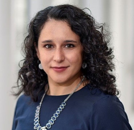
April 11, 2024: Rinki Ratnapriya, Ph.D.
Seminar Title: Using Functional Genomics to Identify the Mechanisms Underlying Age-related Macular Degeneration
Seminar Time: 3 p.m. via Zoom or in-person
Speaker Bio: Dr. Ratnapriya is an assistant professor in the Department of Ophthalmology. She earned her B.Sc. from Delhi University in India and her Ph.D. in Human Genetics from JNCASR, India. She completed a postdoctoral fellowship in Ocular Genomics at the National Eye Institute in Dr. Anand Swaroop’s group, where she utilized next-generation sequencing-based, genome-wide methods to understand the genetic basis of Mendelian and complex neurodegenerative diseases leading to vision loss in humans. She spearheaded a study analyzing the transcriptome and genetics of around 500 donor retinas to establish a reference of expression quantitative trait loci (EyeGEx) to close the knowledge gap between GWAS findings and causal variants, genes, and disease mechanisms. Her research program at BCM focuses on elucidating disease circuitry by extracting valuable information from large genomic, transcriptomic, and epigenetic datasets to address translational and bioinformatic challenges related to retinal and macular degenerative diseases. Additionally, she serves as a faculty mentor for the Data2Knowledge lab at Rice University, facilitating interdisciplinary collaboration among students and faculty to leverage data science for impactful research. She has been recognized with prestigious awards including the NEI Scientific Director’s Award, NEI’s Fellows Award for Research Excellence, David R. Hinton MD Scholarship, and Research to Prevent Blindness Career Development Award. Her research is presently supported by grants from the BrightFocus New Investigator Award, Research to Prevent Blindness Career Development Award, and Foundation Fighting Blindness Individual Investigator Award.
Seminar Summary: In contrast to the unprecedented success in identifying disease risk variants in Age-related Macular Degeneration by Genome-wide association studies, the progress of elucidating the functional relevance of genetic findings in AMD has significantly lagged. This is mainly attributed to our limited understanding of functional impact of associated, non-coding variants on gene function and molecular mechanisms underlying the disease. Trait and disease-associated variants are enriched in the cis-regulatory elements. Thus, gene expression regulation has emerged as a reliable intermediatory between non-coding risk variants and disease phenotype. We focus on elucidating the disease circuitry by extracting valuable information from large genomic, transcriptomic and epigenetic data to connect AMD-GWAS loci to causal variants, target genes and underlying mechanisms. The long-term goal for this research is to leverage the ever-expanding field of data science and genomics to translate the genetic findings in AMD for improving disease diagnosis, management and therapy.
March 21, 2024: Igor Butovich, Ph.D.
Seminar Title: A Concept of Meibogenesis
Seminar Time: 3 p.m. via Zoom or in-person
Speaker Bio: Dr. Igor Butovich is Associate Professor of Ophthalmology in UT Southwestern Medical Center. His research focuses on the causes of, and finding best treatment for, dry eye syndrome.
Seminar Summary: The goal of this seminar "A Concept of Meibogenesis" is to discuss lipid homeostasis of the ocular surface in the norm and pathology. Meibogenesis is defined as an array of Meibomian gland-specific biosynthetic reactions, and corresponding regulatory and signaling mechanisms, that lead to formation of a lipid-rich holocrine secretion called meibum. Meibum is believed to form a protective layer that isolates the surface of the eye from detrimental environmental factors, and improves vision by changing refractive properties of the cornea, though other possible functions of meibum are discussed in the literature as well. Meibum is comprised primarily of extremely long chain and branched compounds that belong to various lipid classes such as wax esters, cholesteryl esters, and a range of other complex lipids. In healthy age- and sex-matched subjects, the lipid composition of meibum is very conservative and changes only minimally from subject to subject. However, a MG pathology called MG dysfunction results in dramatic quantitative and/or qualitative changes in meibum production, negatively affecting the ocular surface physiology, vision, and quality of life in general. MGD is a major contributing factor to a widespread condition called Dry Eye syndrome. The molecular mechanisms of the onset and progression of MGD are currently poorly understood, but believed to be of a multifactorial nature. In this seminar, we will discuss the history and recent advances in the area of MG biochemistry and physiology, as well as major bioanalytical approaches that have been developed to study meibogenesis in human and experimental animals.

Feb. 8, 2024: Yang Sun, M.D., PhD.
Seminar Title: Inositol metabolism and eye diseases
Time: 3 p.m., in-person only. Cullen Eye Institute Auditorium, NC202, 6565 Fannin St. Houston
Speaker Bio: Dr. Yang Sun is a clinician-scientist in ophthalmology and a Professor and Vice Chair, Academic Affairs, of Ophthalmology at Stanford University and Byers Eye Institute. He is the Laurie Kraus Lacob Faculty Scholar at Stanford Child Health Research Institute. Dr. Sun received his BA in Biophysics from Johns Hopkins University, followed by MD and PhD degrees from Washington University School of Medicine. He completed Ophthalmology residency at Stanford University and a prestigious Heed fellowship at University of Michigan, Ann Arbor. Dr. Sun was an Assistant Professor at Indiana University, where he was promoted to an Associate Professor with tenure, before returning to Stanford University. He has been continuously funded by National Eye Institute and Veterans Administration. Dr. Sun’s research in glaucoma has also been funded by American Glaucoma Society, Lowe Syndrome Association, Knights Templar Eye Foundation, and Matilda Ziegler Foundation. Dr. Sun holds several U.S. patents on novel regulators of eye pressure and is the primary investigator on a number of glaucoma clinical trials. He is a member of Stanford BioX faculty and he was elected as a member of American Society of Clinical Investigators.
Seminar Summary: Phosphoinositides are involved in many cellular processes regulating various cellular functions, including signal transduction, membrane trafficking, and cell growth. Enzymes that control phosphoinositide levels are essential for normal development. Mutations in phosphoinositide 5-phosphatases can cause Lowe syndrome, Joubert syndrome, and Marinesco-Sjogren syndrome. We will discuss the role of these 5-phosphatases in congenital glaucoma, cataract, and retinal degeneration.
Jan. 11, 2024: Elizabeth Zuniga-Sanchez, Ph.D.
Seminar Title: Deciphering the developmental mechanisms of retinal circuit assembly
Seminar Time: 3 p.m. via Zoom
Speaker Bio: Dr. Elizabeth Zuniga-Sanchez is an assistant professor in the Department of Ophthalmology and with a joint appointment in the Department of Neuroscience at Baylor College of Medicine.
Seminar Summary: Normal vision relies on a precise number of different neuron subtypes coming together to form a functional circuit. However, the molecular basis of how different neuron subtypes become incorporated into a developing circuit remains understood. Our lab seeks to identify the key molecules responsible for proper neural circuit assembly using the mouse retina as a model system. In the mouse retina, bipolar neurons are interneurons that transmit visual information from photoreceptors to ganglion cells. To date, there are 15 different types of bipolar neurons that differ based on their gene expression profiles, morphology, and connectivity. During development, bipolar neurons are born in excess and through programmed cell death, a specific number of each bipolar subtype remain to give rise to the retinal circuit. Although bipolar development has been well-described, little is known about the molecular mechanisms that mediate bipolar subtype integration. In this seminar, I will present data of how a non-clustered Protocadherin expression code may be responsible for assembling the precise number of each bipolar subtype into a developing circuit. Through this work, we aim to uncover general principles of how different neuron subtypes are assembled during development.
Seminar Speakers 2023

Seminar Title: The neural circuits for light-mediated behavior are more than meets the eye
Time: 3 p.m. via Zoom
Speaker Bio: Dr. Anna Matynia is an Associate Professor in Vision Science at the University of Houston College of Optometry. She received her B.Sc. from Queen’s University in Canada and her Ph.D. in cell biology from Baylor College of Medicine. She completed a postdoctoral fellowship in neuroscience at the University of California, Los Angeles in Dr. Alcino Silva’s group. Dr. Matynia served as research faculty at the University of California, Los Angeles at the Jules Stein Eye Institute where she pioneered research into the neural pathways for photophobia. She currently has an RO1 and U01 grant from NEI, with her research focusing on neural circuits associated with clinically relevant disease or dysfunction in both the retina and cornea. The current funded projects in her laboratory are 1) investigating role of melanopsin in retinal and trigeminal circuitry underlying photo allodynia (light-induced or -enhanced pain), 2) precision mapping of corneal afferents for blink, lacrimation and nociception, and 3) identification of early retinal biomarkers of neurodegeneration in Alzheimer’s Disease.
Seminar Summary: Photoallodynia/photophobia is a clinical condition in which light-induced or -enhanced pain ranges from mild to severely debilitating. The neural pathways mediating photoallodynia were virtually unknown a decade ago. Dr. Matynia will present evidence from clinically relevant models that identifies both retinal and trigeminal contributions to photoallodynia, focusing on the role of melanopsin-expressing neurons. The identification of a specific cell population that contributes to corneal function, raised the question of whether other corneal reflex pathways can be attributed to distinct neural circuits. Dr. Matynia will discuss current and future challenges in identifying these pathways.

Oct. 12, 2023: Melanie Samuel, Ph.D.
Seminar Title: Neurons are not alone: surprising regulators of glia and vessels in the retina
Time: 3 p.m. via Zoom
Speaker Bio: Melanie Samuel is an Associate Professor of Neuroscience and CPRIT Scholar in the Huffington Center on Aging and Department of Neuroscience at Baylor College of Medicine. As a Barry M. Goldwater Scholar, she earned three bachelor’s degrees from the University of Idaho and then completed her Ph.D. at Washington University in neuro-immunology with Michael Diamond. As a postdoctoral fellow with Joshua Sanes at Harvard University she uncovered molecular pathways that determine neural resiliency. Dr. Samuel’s interdisciplinary research group aims to decode the molecular regulators of neuron and glia interaction in order to understand mechanisms that may predispose the brain to diseases. Her past awards include those from the Howard Hughes Medical Institute, the Damon Runyon Cancer Research Foundation, the Brain Research Foundation, a Pathway to Independence Award from the NIH, the NIH Director’s New Innovator Award, and Norton Rose Fulbright Award, a Mallinckrodt Scholar award, and several active R01 awards.
Seminar Summary: The Samuel lab seeks to identify and understand the molecular mechanisms that govern information flow in the nervous system to repair these systems in disease. Much attention has been dedicated to solving the brain’s ‘connectome,’ or street map. Equally important is understanding the road signs and signals that keep traffic (information) moving. This process occurs at synapses. These connections give meaning to the map and underlie all cognition, thought, and behavior. Because nearly all cognitive diseases involve declines in synapse function, deciphering synaptic regulators remains one of the most significant challenges in neuroscience. This talk will focus on surprising non-neuronal regulators of synapses and circuit function, microglia and the vasculature, that are often ignored in the context of wiring studies. Understanding the fundamental processes by which microglia and the vasculature regulate synapses will provide new intervention targets for treating neurological diseases, nearly all of which are accompanied by synapse dysfunction and loss.

Sept.14, 2023: Jijie Pang, M.D., Ph.D.
Seminar Title: Retinal Mechanical Sensation and Scotopic Light Signaling
Time: 3 p.m. via Zoom
Speaker Bio: Dr. Jijie Pang is an associate professor at the Department of Ophthalmology at Baylor College of Medicine. She earned her M.D./Ph.D. degree at Xi'an Jiaotong University, China. Thereafter she completed her postdoctoral training at Baylor College of Medicine. Her work focuses on retinal visual signaling in physiological and pathological conditions. She characterizes retinal photoreceptors, bipolar cells, amacrine cells and ganglion cells with light-evoked activities and single-cell immunofluorescence. Based on such neuronal identities, she further studies the communication between photoreceptors and bipolar cells and synapses among bipolar cells, amacrine cells and ganglion cells, primarily by using whole-cell patch-clamp techniques, in conjunction with genetic engineering, electron microscopy and immunocytology. Built on these physiological data, her study further explores roles of mechanical signals in normal retinal visual pathways and in pathogenesis of glaucoma, as well as other retinal diseases related to physical stresses.
Seminar Summary: In recent decades, a growing number of studies have reported mechanical-sensitive channels (MSCs) in retinal neurons. Our recent studies observed the expression of several types of MSCs in the inner and outer retinal neurons and first reported the pressure-evoked current and voltage response in photoreceptors and bipolar cells. The data have demonstrated that outer and inner retinal neurons are functionally mechanosensitive. Traumatic retinal injury and glaucoma are retinal disorders closely related to pressure. Our current studies funded by DoD focus on the role of MSCs in pressurized air wave-induced traumatic retinal injury. Traumatic retinal injury and glaucoma often impair the peripheral and paracentral retina. Our previous data have identified the glaucomatous eyeball expansion and pressure-induced malfunction in retinal ganglion cells and rod-drive AII amacrine cells. Our recent data generated a novel equation to describe the ocular mechanical hemostasis. Our recent studies attempt to address the function and variation of rod bipolar cells and ganglion cells, the signal integration among multiple photoreceptors in the secondary rod pathway, and a potential novel rod pathway formed by rod bipolar cells. I primarily use patch-clamping recording approaches in these studies, such as the state-of-art dual-cell patch-clamping and the most advanced multi-cell patch-clamp techniques.

Aug. 10, 2023: Mrinalini Hoon, Ph.D.
Seminar Title: The mechanisms underlying assembly of retinal circuits
Time: 3 p.m. via Zoom
Speaker Bio: Dr. Mrinalini Hoon is an Assistant Professor at the University of Wisconsin-Madison Department of Ophthalmology and Visual Sciences. She is also the director of the University of Wisconsin-Madison School of Medicine's electron microscopy facility. She earned her bachelor's degree in Physiology from the Calcutta University and has a PhD in Neuroscience from the International Max Planck Research School at the University of Goettingen, Germany. Thereafter she completed her postdoctoral training with Dr. Rachel Wong at the University of Washington. Her lab is currently funded by grants from NIH/NEI as well as grants from other research foundations. Her research program employs a multi-faceted toolkit to determine the mechanisms that regulate the formation, maturation and maintenance of functional circuits in the mammalian retina.
Seminar Summary: Processing through retinal circuits underlies visual function. Yet we know little about the mechanisms that establish precise connections between retinal cell types. By combining murine transgenic tools with single cell electrophysiology and high-resolution light and electron microscopy we are studying the mechanisms that regulate the establishment of inner retinal synapses specifically the inhibitory synapses at bipolar cell terminals that regulate visual information transfer. We find that a specific early GABA receptor type plays a key role for the establishment and function of these synapses. Additionally, the maturation of these synapses is controlled by visually driven activity. Thus, distinct molecular and activity-dependent mechanisms shape the assembly of retinal microcircuits. Understanding the mechanisms that regulate circuit formation is crucial to determine pathways that are perturbed during disease and degeneration conditions.
July 20, 2023: Rui Chen, Ph.D.
Seminar Title: Single cell multi-omics atlas of the human retina from development to disease
Time: 3 p.m. via Zoom
Speaker Bio: Dr. Rui Chen is Professor of Molecular and Human Genetics, Baylor College of Medicine. He received bachelor’s degree from Tsinghua University, China and PhD degree from Baylor College of Medicine. After completing Postdoctoral training, he joined Department of Molecular and Human Genetics as Assistant Professor, Associate Professor and full Professor. He is the Director of Center of Single Cell Omics at Baylor College of Medicine and has extensive experience in single cell omics profiling.
Seminar Summary: Single cell genomic technologies are revolutionizing basic and translational research in many medical fields, including ophthalmology. I will provide update on the generation of the version 1 single cell atlas of the human retina as part of the human cell atlas (HCA) effort, which includes transcriptomics and open chromatin profile of over 100 cell types from the retina. In addition, findings from single nuclei multi-omics profiling of developing human fetal retina will be discussed. Finally, to facilitate the access of the data source by the community, information of the database and interactive cell atlas browser will be presented.
June 15, 2023: Vivien Coulson-Thomas, Ph.D.
Seminar Title: Hyaluronan (HA) prevents age-related meibomian gland dysfunction
Time: 3 p.m. via Zoom
Speaker Bio: Dr. Vivien J. Coulson- Thomas is an Associate Professor in Vision Science. Dr. Coulson- Thomas completed post-doctoral training in glycosciences and neurobiology at the University of Cambridge in the United Kingdom, and in ophthalmology at the University of Cincinnati. She earned her B.S. in Biomedicine and her MSc and PhD in molecular biology from the Federal University of Sao Paulo. Throughout her education, Dr. Coulson- Thomas earned various awards including the JBC/Herb Tabor Young Investigator Award from the Journal of Biological Chemistry in 2014, British Society of Matrix Biology Young Investigator Award in 2015, the award for excellence in Research, scholarship and creative activity from the University of Houston in 2022, and most recently the Endre Balazs & Janet Denlinger Award from the international society for hyaluronan sciences. She has over 25 years’ experience in the field of glycosciences, and over 15 years’ experience working with the Ocular Surface, with almost 50 published pier reviewed papers. Her research focuses on studying how the extracellular matrix regulates the development, homeostasis and pathology of the ocular surface for which she currently has two RO1 grants from NEI. Most recently, her lab is working on understanding how changes in the extracellular matrix with aging affect dry eye disease.
Seminar Summary: The prevalence of dry eye disease (DED) ranges from ~5 to 50%, with ~85% of all cases being caused by Meibomian gland dysfunction (MGD). As humans and mice age, their Meibomian glands (MGs) undergo age-related changes resulting MG atrophy and dropout, named age related-MGD (ARMGD). The etiology of ARMGD remains elusive, which makes developing therapies to prevent ARMGD extremely challenging. We previously demonstrated that hyaluronan (HA) regulates MG morphogenesis and homeostasis, with an increase in HA leading to an increase in both the number of meibocytes and acini, and, consequently, enlarged glands. We are currently investigating the role of HA in the aging MG, and its therapeutic potential for preventing ARMGD. For such, hyaluronan synthase (Has) knockout mice were aged and compared to age matched wt mice. Our data shows that at 1 year, Has1-/-Has3-/- mice present significantly enlarged MGs, compared to age-matched wt mice and compared to all adult mice. At 2 years, wt mice present severe MG atrophy and dropout, while aged matched Has1-/-Has3-/- mice present healthy glands. As wt mice age, they present a loss of HA surrounding the MG and changes in the composition of the HA matrix, which is correlated with MG atrophy. A loss of HA surrounding the MG in wt mice precedes MG atrophy. Whereas, in contrast, Has1-/-Has3-/- mice present a significant increase in HA deposition, and, consequently do not present MG atrophy. A decrease in HA synthesis in Has1-/-Has3-/- mice, via administration of a chemical inhibitor, is enough to cause MGD. Taken together, our data shows that increasing the HA matrix surrounding the MG can prevent ARMGD in mice.
May 11, 2023: Roxana Radu, M.D.
Seminar Title: What Is the Role of ABCA4 In the Retinal Pigment Epithelium?
Time: 3 p.m. via Zoom
Speaker Bio: Dr. Roxana Radu, M.D., is an Assistant Professor of Ophthalmology at the UCLA Stein Eye Institute and Department of Ophthalmology, David Geffen School of Medicine at UCLA. She possesses extensive education in both clinical and basic-research medical sciences and specific expertise in retinoid biochemistry, which she acquired during her postdoctoral training with Dr. Gabriel Travis. Dr. Radu has established a translational research program that aims to address pressing unmet needs for experimental models of macular degeneration, for which no therapies currently exist. Her group has developed disease models, including mouse lines and human iPSC-derived RPE cultured cells, to elucidate the underlying mechanisms of photoreceptor cell loss in Stargardt disease (STGD1), a juvenile maculopathy, and its relation to age-dependent macular degeneration (AMD). The recent discovery that the ABCA4 gene responsible for STGD1 is expressed and functional in RPE cells has changed the "status quo" of STGD1 pathophysiology. Moreover, Dr. Radu reported for the first time a link between innate immunity dysfunction and STGD1, providing evidence of bisretinoid-mediated complement dysregulation via the alternative pathway in RPE cells carrying an AMD risk allele for the complement factor H (CFH) gene. Current efforts focus on deciphering the intersection of vitamin A-lipid metabolic pathways to understand the biological significance of lipid deposition in RPE cells and identifying key molecular targets for potential therapies for ABCA4-maculopathies.
Seminar Summary: In recent years, the retinal pigment epithelium (RPE) has emerged as a promising cellular target for treating degenerative diseases of the macula. This region supports color vision at high spatial and temporal resolutions, which are essential for many daily activities. Macular degeneration causes the RPE to become dysfunctional and eventually die, leading to photoreceptor degeneration and central visual loss. Currently, there are no therapies for non-exudative macular degenerations such as STGD1 and the dry form of AMD. In this presentation, Dr. Radu will discuss the underlying mechanisms of RPE dysfunction and cell death relevant to STGD1 and AMD. The discovery of ABCA4 expression in RPE cells, in addition to photoreceptors, has led to the development of new models to investigate the RPE cell-autonomous drivers of pathology for these diseases. Dr. Radu will explore the biological processes at the intersection of the complement system, retinoid-lipid metabolism, and mitochondrial bioenergetics and their implications for potential therapeutics.
April 13, 2023: Ross Poché, M.D., Ph.D.
Seminar Title: Awakening the Regenerative Potential of the Mammalian Retina
Time: 3 pm via Zoom
Speaker Bio: Dr. Ross Poché is Associate Professor of Integrative Physiology, Baylor College of Medicine. He received his bachelor degree from the University of New Orleans and PhD from Baylor College of Medicine. His research focuses on transcriptional and epigenetic mechansims regulating retinal progenitor cell proliferation and differentiation.
Seminar Summary: One of my lab's long-term goals is to elucidate the transcriptional and epigenetic mechanisms regulating retinal progenitor cell proliferation, differentiation, and regeneration leading to new therapeutic interventions to restore sight. Toward that end, projects aimed at characterizing mouse mutants suffering from defects in retinogenesis are ongoing. One specific aspect of our research is to identify new strategies to promote Müller glial (MG) cell-mediated retinal regeneration in response to photoreceptor damage. Here, we are interested in determining whether the mouse retina retains latent regenerative potential akin to other vertebrates, such as the zebrafish, and whether we can genetically "awaken" that potential to restore sight. The talk will highlight the discoveries made regarding Hippo pathway regulation of damage-induced Müller glial cell reprogramming to a proliferative, progenitor-like state. I will also present preliminary data describing an approach to isolate native Müller glia and rod proteomes in response to damage.

March 23, 2023: Dong Feng Chen, M.D., Ph.D.
Seminar Title: Harnessing immune balance to combat glaucoma
Time: 3 p.m. via Zoom
Speaker Bio: Dr. Dong Feng Chen, M.D., Ph.D., is Associate Professor of Ophthalmology at the Schepens Eye Research Institute of Massachusetts Eye and Ear and the Department of Ophthalmology, Harvard Medical School. She was trained in the laboratory of Nobel Laureate, Professor Susumu Tonegawa at MIT, before joining the faculty of Harvard Medical School. Her laboratory researches the mechanisms controlling neurodegeneration and regeneration in the eye and brain and they were the first to report full-length optic nerve regeneration from the eye to the brain in genetically engineered post-born mice. Recently, they uncovered an unexpected link among microbiome, autoimmune T cell responses, and neuron and vision loss in glaucoma. These findings received news commentaries in Nature Review Immunology and EyeWorld, etc., highlighted the potential of paradigm shifts in diagnosis, management and treatment of glaucoma and other neurodegenerative diseases in the eye and brain.
Seminar Summary: Glaucoma affects 80 million people worldwide and is a leading cause of irreversible blindness, yet the underlying causes of vision loss are not fully understood. One reason for this is that the eye has traditionally been considered an immune privileged site, which means that there has been little understanding of the involvement of the adaptive immune system, including T cells, in the initiation and progression of the disease. During this presentation, Dr. Chen will describe her team's journey from developing an inducible mouse model of glaucoma to uncovering the role of commensal microbiota-sensitized Th1 cells, as well as the interaction with the innate immune system (specifically microglia), being the root cause of neurodegeneration in glaucoma. Lastly, Dr. Chen will critically discuss the translation studies of their work and the potential implications for the future diagnosis, management, and treatment of glaucoma.
Feb. 9, 2023: Zheng Jiang, Ph.D.
Seminar Title: Spontaneous glial cell activities in healthy and glaucomatous mouse retinas
Time: 3 p.m. via Zoom
Speaker Bio: I have 18 years of experience in retina physiology research. During my doctoral training in Dr. King-Wai Yau’s lab at Johns Hopkins University, I discovered a novel phototransduction pathway in M2- and M4-ipRGCs, which is mediated by cyclic nucleotide and HCN channels. This finding reveals a complex heterogeneity in phototransduction among ipRGCs and, more importantly, breaks a general dogma about segregation of the two phototransduction motifs in the animal kingdom. I joined the Department of Ophthalmology at Baylor College of Medicine as an assistant professor in 2019. My laboratory is focusing on two major directions: i) understanding phototransduction mechanisms and potential applications of melanopsin transduction pathways; ii) studying the electrophysiological properties of retinal ganglion cells in normal and diseased retinas. Along the scientific journey, we discovered a novel electrical activity in retinal glial cells and two new defects in an early-stage glaucoma model.
Seminar Summary: Retinal glial cells play an important role in the maintenance of normal retinal biology. Müller cells are the principal glial cells of the vertebrate retina and span the entire depth of the retina from the outer limiting membrane to the inner limiting membrane. Importantly, despite the critical role of retinal glial cells in supporting normal RGC physiology, it remains unknown whether these cells and/or their interactions with RGCs participate in early-stage glaucoma. Our new approach uses a cutting-edge physiological recording system to monitor extracellular potentials from freshly dissociated retinas. In normal adult mouse retinas, we discovered a glial spontaneous slow voltage fluctuation (SSVF) that has never been reported before. We further demonstrated that the source of SSVFs is likely to be Müller cells. By using a custom algorithm to automatically detect and align these glial and RGC events, we can now efficiently study glial-RGC interactions with spatial and temporal precisions on a large scale. Our preliminary results show a novel glial functional defect in the early stages of a glaucoma mouse model, suggesting that glial cell hyperactivity is a major factor in early glaucoma pathogenesis.
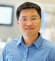
Seminar Title: Glycolysis in retinal angiogenesis and subretinal fibrosis
Time: 3 p.m. via Zoom
Speaker Bio: Dr. Huo is a professor of cellular biology and anatomy, director of the Vascular Inflammatory Program, Vascular Biology Center, Medical College of George, Augusta University. He obtained medical training and served as an internal medicine physician and cardiologist for three years in China. Subsequently, He pursued Ph.D. graduate training at the Medical School of Peking University (China), obtaining expertise in eNOS/nitric oxide biology and endothelial inflammation. He pursued postdoctoral training in Dr. Klaus Ley’s laboratory at the University of Virginia, learning in vivo microscopic approaches to study leukocyte/platelet/vascular interactions in mice. Dr. Huo primary research interests are to study how leukocytes and vascular cells interact to participate in cardiovascular disease and obesity/metabolic disease.
Over past 10 years, Dr. Huo expanded his research to retinopathy field. By collaborating with his colleagues in the James and Jean Culver Vision Discovery Institute at MCG of Augusta University, his group studied the molecular mechanism underlying the role of adenosine receptor 2A (A2aR) and adenosine kinase (ADK) in regulation of ocular angiogenesis and have found that A2aR/adenosine-mediated glycolysis/VEGFR2 in endothelial cells plays a critical role in pathological retinal angiogenesis. His group also demonstrated that PFKFB3-mediated endothelial glycolysis is critical for angiogenesis, especially for the development of oxygen-induced retinopathy (OIR) in mice. Furthermore, his group also revealed that a glycolysis-dictated reciprocal activation between retinal endothelial cells and microglia/macrophages is critical for retinal/choroid angiogenesis. This study was featured in NIH/NEI. These research work reveal an important role of glycolysis in the development of pathological angiogenesis and fibrosis in ocular system.
Seminar Summary: Disorders such as angiogenesis and fibrosis in retinas and choroids involve participation of vascular cells, immune cells as well as mesenchymal cells. For these cells, glycolysis is one of major cellular metabolic pathways. PFKFB3 (6-phosphofructo-2-kinase/fructose-2, 6-bisphosphatase isoform 3) is a critical enzyme for activation of glycolysis in vascular cells and leukocytes. It catalyzes the synthesis of fructose-2,6-bisphosphate (F2, 6P2), which is the most potent allosteric activator of 6-phosphofructo-1-kinase (PFK-1), a rate-limiting enzyme for glycolysis. We generated mice with deficiency of Pfkfb3 in endothelial cells or myeloid cells and used these mice to study how glycolysis in ECs and myeloid cells is involved in pathological angiogenesis and fibrosis in murine models of oxygen induced retinopathy (OIR) and laser induced choroidal neovascularization (CNV). These studies reveal a critical role of glycolytic pathway in the development of these retinal disorders. Additionally, these studies also indicate that inhibiting glycolysis is a promising strategy in the treatment of these diseases.
Seminar Speakers 2022
Seminar Title: Elucidating the molecular mechanisms of neural circuit assembly
Time: 3 p.m. via Zoom
Speaker Bio: Dr. Elizabeth Zuniga-Sanchez is an assistant professor in the Department of Ophthalmology at Baylor College of Medicine. She completed her postdoctoral studies in the Howard Hughes Medical Institute lab of Dr. Larry Zipursky at UCLA where she was one of the first to work on mouse development. In a joint collaboration with the lab of Joshua Sanes from Harvard, they combined two transformational technologies, RNA sequencing of different neuronal subclasses and a rapid CRISPR/Cas9-based genetic scheme to test function of specific genes. Using this approach, they uncovered a new signaling pathway responsible for proper synaptic layer formation. As a postdoctoral fellow, Dr. Zuniga-Sanchez was selected as a Helen Hay Whitney fellow and awarded a K99/R00 Pathway to Independence grant. In her independent research, Dr. Zuniga-Sanchez continues to elucidate novel molecular pathways in neural circuit formation using the mouse retina as a model system. Her research is currently funded by a career development award from Research to Prevent Blindness (RPB), an ARVO Genentech Career Development Award for Underrepresented Minority Emerging Vision Scientists, and an R01 from the National Eye Institute.
Seminar Summary: During the development of the nervous system, a myriad of neuronal cell types assemble into a network to form highly stereotypic patterns of connections. How the dendrites and axons from different neuronal subtypes discriminate between one another to form precise and highly reproducible patterns of synaptic connections remains a central issue in neuroscience. Our research focuses on identifying the molecular basis of neural circuit assembly in the developing vertebrate nervous system. We use single cell sequencing, CRISPR technology, live imaging, and single cell labeling to identify the factors that mediate proper connectivity using the mouse retina as a model system. Through these studies, we aim to uncover general principles of neural circuit formation in the developing mammalian nervous system.
Oct. 11, 2022: Stephen Pflugfelder, M.D.
Seminar Title: Retinoid modulation of ocular surface inflammation
Time: 3 pm via Zoom
Speaker Bio: Stephen C. Pflugfelder, M.D., is professor and holder of the James and Margaret Elkins Chair and director of the Ocular Surface Center in the department of ophthalmology at Baylor College of Medicine. He specializes in cornea, ocular surface and tear disorders. Dr. Pflugfelder’s research interests include pathogenesis of keratoconjunctivitis sicca and desiccation-induced inflammation and autoimmunity on the ocular surface. He has published more than 340 peer-reviewed articles and numerous book chapters. He is past president of the International Ocular Surface Society and has served on the editorial boards of Investigative Ophthalmology and Visual Science (associate editor), American Journal of Ophthalmology (associate editor), Scientific Reports, Cornea and The Ocular Surface. He served on the ARVO board as Cornea Trustee and President. He is a member of the Department of Defense Vision Research Panel.
Seminar Summary: Dry eye stress stimulates innate inflammatory pathways in ocular surface epithelial and immune cells. Myeloid cells are recruited to the conjunctiva that produce inflammatory mediators that sensitize nociceptors and stimulate IL-17 production by gamma and delta T cells. IL-17 promotes corneal barrier disruption and conjunctival cornification and goblet cell loss. Retinoid signaling through RXR-alpha suppresses myeloid and gamma delta T cell activation and IL-17 production. Conjunctival and lacrimal gland disease in dry eye can disturb the retinoid axis. The RXR-alpha pathway can be leveraged to treat dry eye inflammation.

Seminar Title: Light-induced neuronal activity drives the initiation of optic pathway glioma in NF1
Time: 3 p.m. via Zoom
Speaker Bio: Dr. Yuan Pan is an assistant professor in the Department of Symptom Research at the University of Texas MD Anderson Cancer Center. Dr. Pan received her Ph.D. degree from the University of Iowa and her thesis work focused on understanding membrane protein trafficking in photoreceptors. As a postdoctoral fellow, Dr. Pan is co-mentored by Dr. David Gutmann (Washington University) and Michelle Monje (Stanford University) and her research focused on optic pathway gliomas. Leveraging genetically engineered mouse models and neuromodulatory approaches, Dr. Pan’s group is currently working on understanding neuron-glia and neuron-cancer interactions, with a focus on cancer predisposition syndromes such as neurofibromatosis type 1 (NF1).
Seminar Summary: Neurons have recently emerged as key players in driving cancer pathogenesis. While the role of neuronal activity in tumor growth is established, the importance of neuronal activity to tumor initiation was less clear. Individuals with the NF1 cancer predisposition syndrome often develop tumors in the optic pathway (optic glioma) during early childhood, raising the possibility that postnatal light-induced optic nerve neuronal activity drives tumor initiation. In this seminar, Dr. Pan will present how she and her colleagues leveraged a unique mouse model of Nf1 optic glioma to identify how light-induced neuronal activity drives tumorigenesis in the optic nerve.
Aug. 9, 2022: Bo Chen, Ph.D.
Seminar Title: Protecting Ganglion Cells and Reprogramming Müller Glia in the Retina
Time: 3 p.m. via Zoom
Speaker Bio: Dr. Bo Chen is professor of ophthalmology and neuroscience at Icahn School of Medicine at Mount Sinai. He received his Ph.D. in pharmacology at the University of Miami School of Medicine, and later pursued postdoctoral training in the Department of Genetics at Harvard University. He was previously an assistant and associate professor in the Departments of Ophthalmology and Neuroscience at Yale University School of Medicine, and is currently a professor in the Departments of Ophthalmology and Neuroscience at the Icahn School of Medicine at Mount Sinai. He received the Karl Kirchgessner Foundation Award for Retinal Research and was a Pew Scholar in the Biomedical Sciences from 2013-2017. He is a reviewer for numerous publications including, but not limited, to Science, Neuron, eLife, PNAS, Cell Reports, Science Advances, and Journal of Neuroscience. Dr. Bo Chen’s research focuses on mechanistic and therapeutic studies of retinal degenerative diseases caused by loss of photoreceptors or retinal ganglion cells, such as age-related macular degeneration, retinitis pigmentosa, and glaucoma. To study these conditions, his laboratory pursues two main strategies: neuroprotective strategy to save existing retinal neurons and neural regenerative strategy to produce new retinal neurons.
Seminar Summary: Retinal ganglion cells (RGCs) and photoreceptors are particularly vulnerable in major retinal degenerative diseases leading to vision impairment and blindness. We will present our recent research progress, as well as future challenges, in protecting RGCs for vision preservation and regenerating photoreceptors by reprogramming Müller glia for vision restoration. Using multiple injury and disease models, we will examine several signaling pathways and transcription factors that may play an important role in the survival and regeneration of RGCs and photoreceptors.
Seminar Title: Glaucoma in mice: new tools, new insights, new questions
Time: 3 p.m. via Zoom
Speaker Bio: Benjamin J. Frankfort, M.D., Ph.D., is an Associate Professor in the Departments of Ophthalmology and Neuroscience at Baylor College of Medicine. He was an undergraduate at Duke University and received his M.D. and Ph.D. degrees from Baylor College of Medicine. He completed a residency in Ophthalmology at the Wilmer Eye Institute of the Johns Hopkins School of Medicine and a fellowship in glaucoma surgery at the Cullen Eye Institute of Baylor College of Medicine. He continued his research training as a K08 recipient under the supervision of Dr. Sam Wu at Baylor College of Medicine. Dr. Frankfort’s research focuses on the impact of eye pressure changes on the visual system. His lab uses a variety of techniques to assess anatomic, physiologic, and molecular changes in injured rodent tissue with resolution ranging from single cells to whole organs. Dr. Frankfort currently holds an R01 and R21 from the National Eye Institute and maintains funding from several foundations. As faculty at Baylor College of Medicine, Dr. Frankfort is highly active in the education of medical students, graduate students, residents, clinical fellows and post-doctoral fellows, and is the Co-Director of the Medical Scientist Training Program (MSTP).
Seminar Summary: Glaucoma is a progressive neurodegenerative disease of retinal ganglion cells (RGCs) and the optic nerve. Despite its wide prevalence and importance as a major public health issue, the earliest changes that occur in glaucoma remain a mystery, hindering early diagnosis and treatment options. In the later stages of glaucoma, RGC regeneration, optic nerve regrowth, and vision restoration techniques are in their infancy.
We will present new techniques developed in the Frankfort lab to study both early glaucoma phenotypes and RGC regeneration. Using the bead injection model of intraocular pressure (IOP) elevation in conjunction with a new vascular assessment workflow, we determined that retinal capillary plexi are differentially susceptible to elevated IOP. Using a series of improvements to RGC immunopanning techniques and optimized FACS, we established a novel system of primary RGC isolation from adult mouse retinas with subsequent culture. With it, we observed robust neurite outgrowth which occurs according to several stereotypical stages.
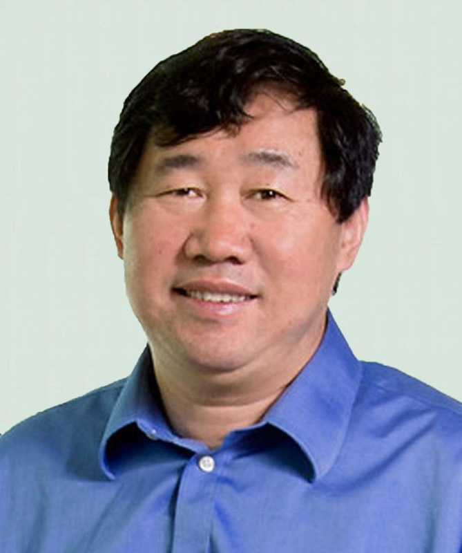
Seminar Title: Deciding a fate: How key transcription factors interact with the epigenetic landscape to promote retinal ganglion cell genesis
Time: 3 p.m. via Zoom
Speaker Bio: Dr. Xiuqian Mu is professor of ophthalmology at the University of Buffalo, Jacobs School of Medicine and Biomedical Sciences. He received his M.D. degree from Qingdao Medical College, China, and his Ph.D. degree in biochemistry and molecular biology from Peking Union Medical College. He received training as a visiting fellow in NIH and postdoctoral associate in MD Anderson Cancer Center, the University of Texas. He was promoted to instructor and assistant professor at MD Anderson Cancer Center and subsequently joined the University of Buffalo as assistant, associate and full professor.
Seminar Summary: My lab is interested in the fundamental mechanisms regulating the shift of cellular states in retinal cell differentiation. One major focus is on how transcription factors influence the epigenetic landscape to promote the genesis of individual retinal cell types such as retinal ganglion cells (RGCs). During development, all retinal cell types emerge from multipotent retinal progenitor cells (RPCs) following distinct trajectories. Recent scRNA-seq studies have further delineated these trajectories, providing a general picture of their relationships and revealing a transitional RPC state shared by all the early lineages. We have now further dissected the changes in the epigenetic landscape along these trajectories by scATAC-seq and identified globally the enhancers, enriched motifs, and potential interacting transcription factors underlying the cell state-specific gene expression in individual lineages. Using CUT&Tag-seq, we identified the enhancers bound directly by four key transcription factors, Otx2, Atoh7, Pou4f2, and Isl1, and uncovered their roles in shaping the epigenetic landscape and controlling the gene expression program in the RGC lineage. Our findings provide experimental support for a general paradigm in which transcription factors collaborate and compete in a sequential and combinatorial fashion to regulate the emergence of distinct retinal cell types such as RGCs from the multipotent RPCs.
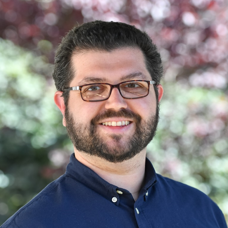
Seminar Title: Selective synaptic disassembly and reassembly of the mature retina in mouse models of glaucoma
Time: 3 p.m. via Zoom
Speaker Bio: Dr. Della Santina is an assistant professor of vision sciences at the University of Houston College of Optometry. He received his Ph.D. in neuroscience from the University of Pisa in Italy studying the functional role of retinal voltage-gated ion channels in processing visual stimuli. He then completed his postdoctoral training at the University of Washington in Seattle under the guidance of Dr. Rachel Wong. His postdoctoral work elucidated the role of neurotransmission in the development of retinal synaptic specificity and identified the relative susceptibility of retinal ganglion cell types to intraocular pressure elevation in mouse models of glaucoma. First at the University of California San Francisco, and now at the University of Houston, his laboratory utilizes electrophysiology, machine learning and large-scale imaging of neural circuits to investigate the effect of retinal neurodegenerative diseases on neural connectivity and functionality.
Seminar Summary: The visual system encodes salient features of the visual scene by employing parallel signal processing pathways. The anatomical basis to such variety starts in the retina with the specific connectivity between types of neurons established through development. The maintenance of this architecture is challenged by neurodegenerative diseases such as glaucoma where post-synaptic neurons, retinal ganglion cells (RGCs) die over time, forcing the existing neural circuits to adapt to a novel environment. We induced RGCs death in mice by experimentally increasing the intraocular pressure and analyzed the intraretinal connectivity of different RGC types with their presynaptic partners, to identify patterns of synaptic rearrangements. We observed that although presynaptic neurons do not die, they lose synaptic ribbons and different RGC types alter their specific connectivity pattern over time and form new connections with former developmental partners. Together, our current data suggests that different neuronal types engage diverse strategies in response to degeneration; leveraging these differences could be the key to develop early functional tests for neurodegenerative diseases.

Seminar Title: Ultrastructural analysis of Cep290 mutant cilia in mouse photoreceptors
Time: 3 p.m. via Zoom
Speaker Bio: Dr. Abigail Moye is a postdoctoral fellow in Dr. Theodore Wensel’s Laboratory, Department of Biochemistry and Molecular Biology, Baylor College of Medicine. Her research is funded by NIH F32 Postdoctoral Fellowship.
Seminar Summary: CEP290 (centrosomal protein 290), a large multi-domain containing protein, is a key component of the transition zone in many primary and motile cilia. Mutations in Cep290 cause several ciliopathies, including Meckel Syndrome (MKS) and retinal specific disorders such as Leber Congenital Amaurosis (LCA). The CEP290 protein has been proposed as a component of the “Y-links” structures, which span from the axoneme microtubule doublets to the ciliary membrane in the connecting cilium (CC) of photoreceptor cells. In this presentation, I will discuss the retinal phenotypes in two Cep290 mutant mouse models, as well as our use of multiple advanced microscopies to determine the location of CEP290 protein and the structural effects resulting from mutations in CEP290 at the nanometer scale.
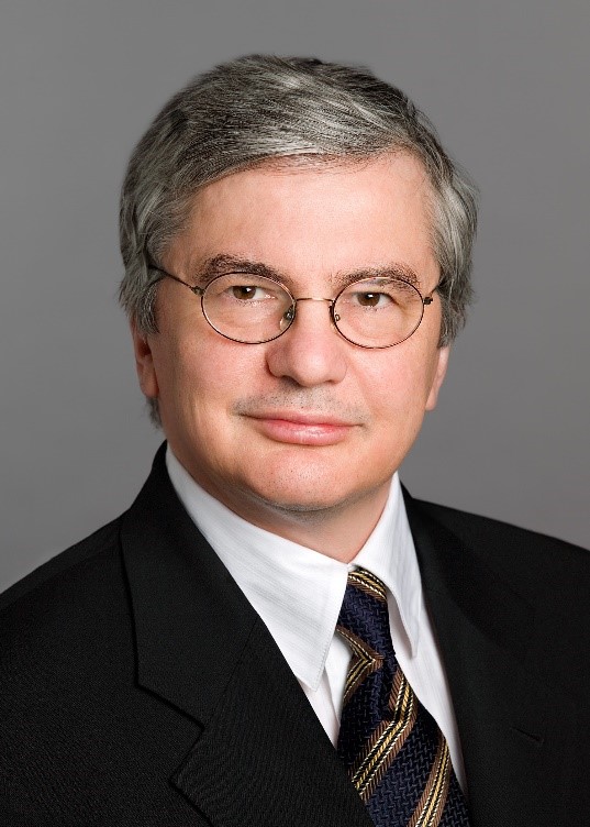
Seminar Title: Identification of Novel Mechanisms of Angiogenesis
Time: 3 p.m. via Zoom
Speaker Bio: Dr. Napoleone Ferrara is distinguished professor of pathology and ophthalmology and Hildyard Endowed chair at UC San Diego, Calif. Before joining UCSD in 2013, he was senior scientist, staff scientist and Genentech fellow at Genentech. At Genentech, he discovered vascular endothelial growth factor (VEGF) and its receptors and developed the first anti-VEGF antibody drug bevacizumab (Avastin) for cancer therapy. His work also led to the development of ranibizumab (Lucentis) for the therapy of ocular vascular diseases. He has received numerous awards, including Lasker-DeBakey Clinical Medical Research Award in 2010 and Breakthrough Prize in Life Sciences in 2013. At UCSD, his research focuses on investigating angiogenic mechanisms in tumor and ocular disease.
Seminar Summary: Dr. Ferrara’s research focuses on investigating mechanisms of angiogenesis in disease conditions alternative to VEGF. He will present his recent findings in therapeutic angiogenesis.

Seminar Title: Top-down feedback controls neural responses in primate visual cortex
Seminar Time: 3 p.m.
Speaker Bio: Dr. Lauri Nurminen is assistant professor of vision science at the University of Houston College of Optometry. He received his Ph.D. degree in neuroscience from the University of Helsinki, Finland. He completed postdoctoral training at the Moran Eye Center of the University of Utah under the guidance of Dr. Alessandra Angelucci. His postdoctoral work elucidated the neural circuit basis of receptive fields of primary visual cortex neurons in non-human primates. Part of his NIH K99 training was completed under the guidance of Dr. John Reynolds at the Salk Institute for Biological Studies where he worked with awake, behaving marmoset monkeys. His laboratory utilizes electrophysiology, optogenetics, and behavioral techniques in awake and behaving marmosets to elucidate the roles of recurrent connections between cortical areas in visual computation and perception.
Seminar Summary: Sensory information travels along feedforward connections through a hierarchy of cortical areas, which, in turn, send feedback connections to lower-order areas. Feedback has been implicated in attention, expectation, and sensory context, but how feedback could implement such diverse functions remains unknown. In my presentation, I will show how we have used optogenetic inactivation of the axon terminals of feedback connections from the second visual area (V2) to the primary visual cortex (V1) in non-human primates (Common Marmoset) to determine how feedback affects neural responses in V1. Our results show that reducing feedback activity increases V1 cells’ receptive field size, decreases their responses to stimuli confined to the RF, and increases their responses to stimuli extending into the proximal surround, therefore reducing surround suppression. Moreover, a stronger reduction of V2 feedback activity leads to a progressive increase in RF size and a decrease in response amplitude. Our results indicate that feedback modulates RF size, surround suppression, and response amplitude, similar to the modulatory effects of visual-spatial attention.
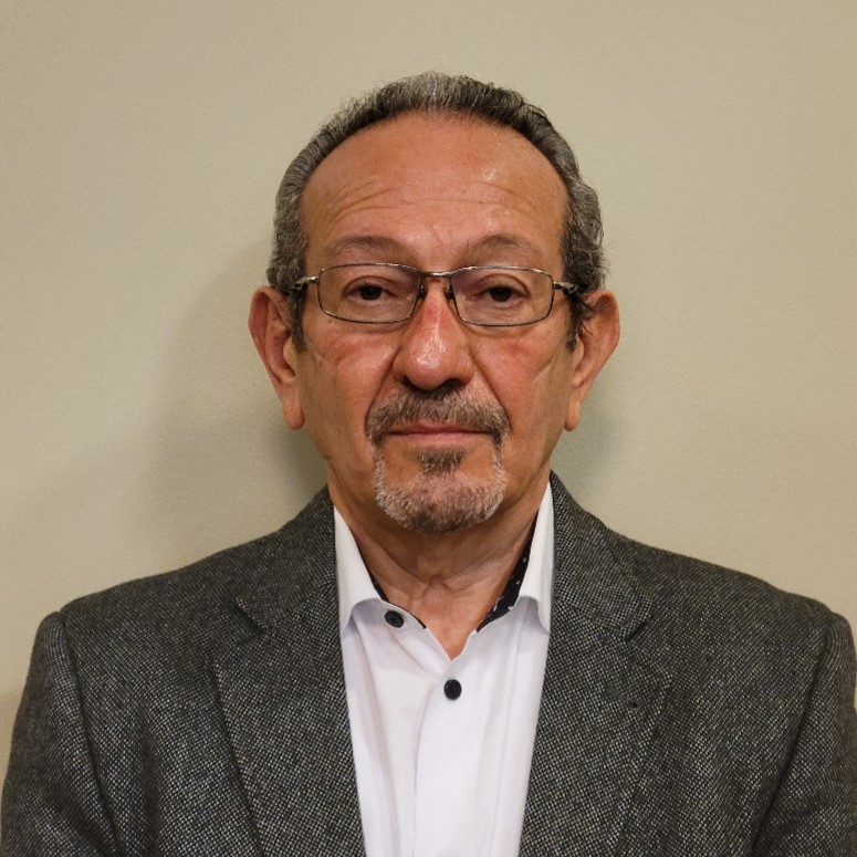
Seminar Title: Role of riboflavin in vision
Seminar Time: 3 p.m. via Zoom
Speaker Bio: Dr. Muayyad Al-Ubaidi is Professor of Biomedical Engineering at Cullen College of Engineering, University of Houston. He is a leader in mouse transgenesis and has been very active in the production of transgenic models of inherited retinal disease.
Seminar Summary: Ariboflavinosis is a pathological condition occurring because of riboflavin (vitamin B2) deficiency. Riboflavin is intracellularly converted to flavin mononucleotide (FMN) and flavin adenine dinucleotide (FAD), both are cofactors for many enzymatic reaction. Ariboflavinosis can be the result of mutation is riboflavin transporters or improper dietary intake of the vitamin. Clinical manifestations of ariboflavinosis include neurological and visual abnormalities. However, the mechanism of the visual defects is not understood. In this presentation, we will address how riboflavin is delivered to the retina, maintained at high level, the mechanism that leads to visual defects resulting from ariboflavinosis and the metabolic changes associated with manipulations in retinal flavin levels.
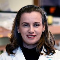
Seminar Title: Investigating IL-2 Corneal Epithelial Signaling
Seminar Time: 3 p.m. via Zoom
Speaker Bio: Dr. Cintia De Paiva is Associate Professor of Ophthalmology at Baylor College of Medicine. Her research focuses on pathogenesis of dry eye-related diseases with the ultimate goal to improve the diagnosis, prognosis and therapy of dry eye.
Seminar Summary: The cornea is richly innervated and responds to various chemical, mechanical, and thermal stimuli to initiate reflex blinking and tearing. Neuropeptides produced by corneal nerves maintain epithelial integrity and stimulate wound healing. In turn, corneal epithelial cells support the nerves functioning as surrogate glial cells. However, the superficial location of the corneal nerves renders them susceptible to desiccating stress, mechanical trauma, and inflammation that can sensitize them, causing pain and excessive blinking. Diseases that affect corneal nerves such as dry eye and neurotrophic keratitis can lead to debilitating ocular pain, ulceration, and opacification and may require aggressive therapies, including corneal transplantation, to restore vision.
IL-2 is a cytokine with a central role in the immune system, promoting activation, clonal expansion, and deletion of T lymphocytes. IL-2 is detectable in the tears of healthy subjects. IL-2 signals through its heterotrimeric receptor (IL-2R) consisting of the alpha (CD25), beta, and gamma chains. IL-2 binds directly to CD25, making this receptor subunit key to IL-2 signaling. Non-immune CD25 and IL-2Rbeta expression have been reported in several different types of epithelial cells. We and others have reported that the corneal epithelium exhibits strong immunoreactivity to CD25. We have provocative and exciting data suggesting that the corneal CD25 is a functional receptor and that an IL-2 corneal epithelial signaling exists.
Seminar Speakers 2021
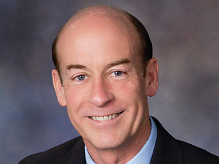
Seminar Title: Retinal Phosphoinositides
Time: 3 p.m. via Zoom
Speaker Bio: Dr. Theodore Wensel is the Robert A. Welch Chair in Chemistry, chair of biochemistry at Baylor College of Medicine. His research focuses on vision research, including visual transduction, G protein-coupled receptors, phosphoinositide signaling, RGS proteins, biomembranes, ocular proteomics and gene repair.
Seminar Summary: Phosphoinositides, the phosphorylated forms of the membrane lipid phosphatidylinositol, play important roles in signal transduction and membrane trafficking. We have found that in rod cells, light exposure leads to dramatic increases in levels of phosphatidylinositol-3-phosphate (PI(3)P) and phosphatidylinositol-4,5-bisphosphate (PI(4,5)P2). We have knocked out the gene (Vps34 aka Pik3c3) encoding the Type III PI-kinase responsible for synthesis of PI(3)P in rods, cones, ON-bipolar cells and RPE cells. In all three cell types, development proceeds normally, but there are defects in autophagy, endosome processing and phagocytosis that lead to cell death. Surprisingly PI(3)P is not required for initiation of autophagy or phagocytosis, but is essential for fusion with lysosomes. PI(4,5)P2 is found in discrete patches in the plasma membrane of rod outer segments, rod inner segments, the outer nuclear layer, and peri-synaptic membranes. Its levels in the light are reduced in the absence of phototransduction, but the mechanism and functions of the light-induced changes remain under investigation. The type I PI-3 kinase responsible for synthesis of PI(3,4,5)P2 does not appear to be important for rod function or survival.
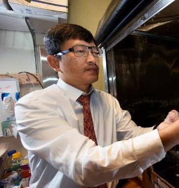
Seminar Title: The role of trophoblast glycoprotein in the retina
Time: 3 p.m. via Zoom
Speaker Bio: Dr. Ching-Kang Jason Chen is professor of ophthalmology and Alice R. McPherson Retina Research Foundation Endowed chair at Baylor College of Medicine. His research focuses on heterotrimeric G-proteins in vision, mechanisms and consequences of photoreceptor degeneration, retinal cell biology and circuitry and phototransduction.
Seminar Summary: Trophoblast glycoprotein (TPBG), recognized by a monoclonal antibody 5T4, is highly expressed in embryonic tissues and restricted in adults. In the retina, it is expressed specifically in rod bipolar cells and in an ON-OFF wide-field amacrine cell called TPBG-AC. We have made and characterized the Tpbg-/- mice and found in them a unique enhancement of photopic contrast sensitivity to certain spatial stimuli. This talk will cover our current working hypothesis and progress on the roles of TPBG in the retina.

Seminar Title: Population-specific mechanisms of retinal ganglion cell neuroprotection and axon regeneration
Time: 3 p.m. via Zoom
Speaker Bio: Dr. Nicholas Tran is an assistant professor of molecular and human genetics, Baylor College of Medicine, Houston, Texas.
Seminar Summary: Neurons in the CNS have limited ability to recover from axonal injury. Much focus has been placed on identifying strategies to protect damaged neurons and stimulate regrowth of axons. While many treatments have been identified, each typically only protects a fraction of neurons and fewer still regenerate their axons. Why do some neurons respond to treatment while others do not? We are applying single-cell transcriptomic approaches to determine how different populations of retinal ganglion cells (RGCs) are affected by injury and how they respond to different neuroprotective and regenerative interventions. We identify population-specific expression programs correlating with RGCs that are likely to degenerate, survive but not regenerate, or survive and regenerate. This work reveals potential gene targets for advancing the development of therapies of both optic neuropathies and acute nerve injuries like spinal cord injury.

Seminar Title: Combating drug resistance in age-related macular degeneration
Time: 3 p.m. via Zoom
Speaker Bio: Dr. Yingbin Fu is an associate professor of ophthalmology and Sarah Campbell Blaffer Endowed chair at Baylor College of Medicine, Houston, Texas.
Seminar Summary: Choroidal neovascularization (CNV), which accounts for 10-20 percent of AMD, is responsible for 80-90 percent of AMD-related blindness. Anti-VEGF therapies that target extracellular VEGF have revolutionized the treatment for CNV; however, it is estimated that up to 50 percent of patients have poor responses to this treatment. Different strategies have been tested to overcome this issue. However, no major breakthrough has been reported in combating anti-VEGF resistance. The mechanisms for anti-VEGF resistance are poorly understood. We explore the unique property of the apolipoprotein A-I (apoA-I) binding protein (AIBP) that enhances cholesterol efflux from endothelial cells and macrophages to thereby limit angiogenesis and inflammation to tackle anti-VEGF resistance in CNV. We show that laser-induced CNV in mice with increased age showed increased resistance to anti-VEGF treatment, which correlates with increased lipid accumulation in macrophages. The combination of AIBP/apoA-I and anti-VEGF treatment overcomes anti-VEGF resistance and effectively suppresses CNV. Furthermore, macrophage depletion in old mice restores CNV sensitivity to anti-VEGF treatment and blunts the synergistic effect of combination therapy. These results suggest that cholesterol-laden macrophages play a critical role in inducing anti-VEGF resistance in CNV. Combination therapy by neutralizing VEGF and enhancing cholesterol removal from macrophages is a promising strategy to combat anti-VEGF resistance in CNV.

Seminar Title: A paradoxical increase in visual sensitivity in a mouse model of retinal degeneration
Time: 3 p.m. via Zoom
Speaker Bio: Dr. Eduardo Solessio is associate professor of ophthalmology and visual sciences and neurosciences and physiology at SUNY Upstate Medical University, Syracuse, NY.
Seminar Summary: Various retinal degenerative diseases in humans minimally cause loss of retinal function, but can also cause a loss of retinal cells, and eventual blindness. In order to improve therapeutic strategies and treatment outcomes for these diseases, it is critical to understand the fundamental retinal and visual changes that occur early in retinal degenerative disease. In this presentation, I will use a mouse model of a human retinal disease to demonstrate an aspect that may develop during early retinal degeneration: an enhanced ability to detect flickering lights. I will also show that this surprising retinal gain-of-function is caused by a pathological acceleration of the temporal properties of rod photoresponses. Advanced rod dysfunction in human disease is currently diagnosed using relatively complicated tests employing full-field ERG and perimetry. Applying our method to humans to measure retinal or visual sensitivity to flickering lights could prove useful as a rapid test for early rod dysfunction in some forms of retinal degeneration.
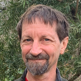
Seminar Title: Deciphering Retinal Cell Fate Determination with Single Cell Omics
Time: 3 p.m. via Zoom
Speaker Bio: Dr. Graeme Mardon is James R. Davis chair in pathology and immunology, professor of molecular and human genetics at Baylor College of Medicine.
Seminar Summary: The Drosophila eye has been one of the most powerful animal model systems for deciphering molecular mechanisms of cell fate determination and development for more than 40 years. A landmark discovery reported in 1996 (Freeman) revealed that the Epidermal Growth Factor Receptor (EGFR) signaling pathway is both necessary and sufficient to trigger the differentiation of nearly all cell types in the Drosophila retina. This finding posed the question of how activation of a single pathway could trigger the differentiation of more than ten different cell types in the same tissue at the same time during development. More than a quarter century later, there has been little mechanistic progress in our understanding of this process. Given the fundamental importance of EGFR signaling throughout higher eukaryotes in cell differentiation, proliferation, and survival, it is of broad significance to unravel this conundrum. To this end, we have generated extensive single cell RNA-sequencing data on multiple stages of the developing Drosophila eye, as well single cell chromatin accessibility data (ATAC-seq). Integration of these data should lead to a better understanding of how the EGFR pathway is reiteratively used to regulate retinal cell fate determination.
Seminar Title: Vision for escape and pursuit
Time: 3 p.m. via Zoom
Speaker Bio: Dr. Daniel Kerschensteiner is professor in Department of Ophthalmology and Visual Science at Washington. University.
Seminar Summary: One interest of my lab to understand how the visual system detects and tracks salient stimuli in the environment to initiate and guide specific behaviors (i.e., visual neuroethology). Predator avoidance and prey capture are central selection pressures of animal evolution. Mice use vision to detect aerial predators and hunt insects. I will discuss studies from my group that identify specific circuits and pathways in the early visual system (i.e., the retina and its subcortical targets) mediating predator avoidance and prey capture in mice. Our results highlight the importance of subcellular visual processing in the retina and the alignment of viewing strategies with region- and cell-type-specific retinal ganglion cell projection patterns to the brain.
Seminar Title: T-box Transcription factors in the development of retinal neuronal subtypes
Time: 3 p.m. via Zoom
Speaker Bio: Dr. Chai-An Mao is assistant professor in Ruiz Department of Ophthalmology and Visual Science at the University of Texas McGovern Medical School.
Seminar Summary: We are interested in understanding the fundamental mechanisms regulating how diverse neuronal types in the central nervous system arise during development. In a mature mammalian retina, more than forty distinct types of retinal ganglion cells (RGCs) and sixty types of amacrine cells (ACs) have been described on the basis of their morphologies, functions, and molecular profiles. However, the cellular and molecular mechanisms that control the specification and differentiation of these diverse neurons remain unclear. This talk will discuss the roles of two evolutionarily conserved T-box containing transcription factors, Tbr1 and Tbr2, in the development of distinct subsets of RGCs and ACs.

Seminar Title: Sidestepping stem cells to restore sight
Time: 3 p.m. via Zoom
Speaker Bio: Dr. Chavala currently serves as a professor in the Department of Surgery at the Texas Christian University/University of North Texas Health Science Center School of Medicine in Fort Worth, Texas.
Seminar Summary: Up until now, researchers have replaced dying photoreceptors in animal models by creating stem cells from skin or blood cells, programming those stem cells to become photoreceptors, which are then transplanted into the back of the eye. Dr. Chavala’s team shows that it is possible to skip the stem-cell intermediary step and directly reprogram skins cells into photoreceptors for transplantation into the retina. The lab-made photoreceptors enabled blind mice to detect light after the cells were transplanted into the animals' eyes. The work was published April 15 in Nature. This is the first study to show that direct, chemical reprogramming can produce retinal-like cells. It provides a novel and faster strategy for developing therapies for retinal disorders caused by the loss of photoreceptors